A Review of Gold Nanoparticle Use in Unique Medical Applications: Combining Anti-bacterial and Anti-cancer Treatment in One Nanoparticle
published: 20 November 2025 | https://doi.org/10.63174/xdi.POJG1530
Abstract
Gold nanoparticles have been studied extensively for various medical applications due to their strong capability for cancer treatment without drugs and their equally strong, but newly identified, potential in the anti-bacterial field. Due to their ease of synthesis, chemical stability, activation by light, and controllable toxicity, gold nanomaterials have attracted enormous interest as novel biomaterials, especially for cancer patients who have an increased rate of infection due to a compromised immune system. This review discussed how gold nanoparticle used in the anti-bacterial and anti-cancer fields, as well as their prospect in further medical research to combine such multi-properties into one kind nanoparticle. Given the adverse impacts of the pandemic spanning 2020 to 2022, a considerable number of research projects were either interrupted or prematurely terminated. As a direct consequence, this review solely encompasses the research progress attained prior to the onset of the pandemic. Notably, the core unresolved issues within this research domain—with specific focus on topics centered around gold nanoparticles—continue to be the subject of ongoing investigation to the present day.
1. Background and Introduction
In 2018, the Global Health Organization reported over three million deaths caused by infection-related diseases. Particularly, lower respiratory infections have become the most deadly communicable diseases across the world.[1] In the last ten years, there has been insufficient clinical trials and research (as well as regulatory approval of new antibiotics), leading to a standstill in new antibiotic development[2, 3]. On the other hand, the emergence of antibiotic resistant bacteria has significantly weakened the efficiency of traditional antibiotics during infection treatment. More critically, it is predicted that antibiotic resistant bacteria will remain (if not grow) at the same level in the perennial future due to the long-time abuse of antibiotics.4 With a pessimistic prediction of antibiotic effectiveness in the foreseeable future, humans are facing a critical situation in the war with bacteria.
Under these circumstances, nanoparticles have become a potential antimicrobial strategy with revolutionary novel anti-bacterial strategies for killing bacterial, aiding the immune system, altering bacteria gene expression, development of bacteria-infecting viruses, and so much more that could become the future of anti-bacterial medicine.[5-8] Compared to traditional antimicrobial strategies using pharmaceutical agents, nanoparticles have less of a development path and compared to bulk materials, its nanostructure inherently provides more anti-bacterial potential due to higher surface to volume ratios.
Another major health concern still plaguing today’s medicine is cancer where in 2018, it was estimated that over 1.8 million people were diagnosed with cancer and over 600 thousand people died from cancer within the United States alone.[9] A similar dilemma to antibiotic resistant bacteria has occurred in the anti-cancer field: a significant increase in multidrug resistant cancer cells as well as a lack of development of novel treatments that do not involve chemotherapeutics.[9-14] It is clear that our current global healthcare system relying on drugs to both kill bacteria and cancer cells has reached its limit.
On the other hand, infection and cancer-related diseases are not always independent issues. Both theoretical and epidemiological statistics support that both virus and bacterial infections are extrinsic risk factors, playing a significant role in the development of some specific cancers.[15-17] Especially for long-term bacterial infectious diseases, bacteria can induce cancer formation, and the two types of cells (bacteria and cancer cells) act in a symbiotic manner especially in a weakened immune system of a cancer patient.[18] For example, there are at least six kinds of cancers that have been found to be bacterial infection related. Specifically, Helicobacter pylori, Salmonella Typhi, Salmonella Enteritidis, and Chlamydia trachomatis promote at least six kinds of cancer, which include non-cardiac gastric carcinoma, low-grade B-cell MALT gastric lymphoma, Gallbladder carcinoma, colon carcinoma in the ascending and transverse parts of the colon, as well as carcinoma of the cervix and ovaries. [19-23] Moreover, clinical research has indicated that cancer patients possess more risk for bacterial infection because of their immunocompromised health. [24, 25] Thus, significant advances in medicine can be made to develop nanoparticles (or any strategy for that matter) that can simultaneously possess anti-bacterial and anti-cancer properties. Figure 1 shows the correlation between bacterial infection and cancer important for the next generation of technologies to treat simultaneously.
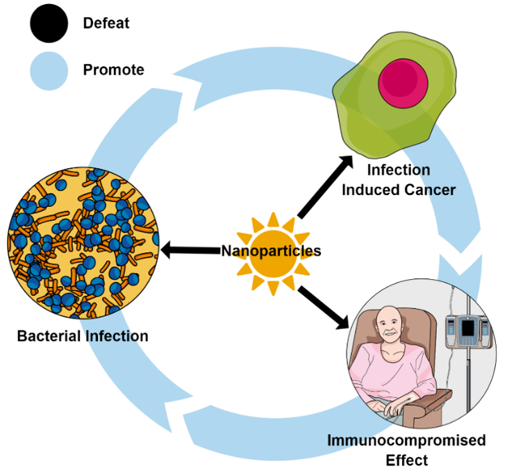
Figure 1. Correlation schematic of bacterial infection and cancer.
Past research has identified several nanoparticles (such as silver, tellurium, and selenium) that have potential capability in both anti-cancer and anti-bacterial fields. [26-29] Similar to other nanoparticles, gold nanoparticles have an exceptional capacity for biological applications, such as their use in the visualization of diseases, drug delivery, photodynamic therapy, bio-sensor, infectious bacteria inhibition, and cancer treatment applications. [30] Due to their ease of synthesis, chemical stability, activation by light, and controllable toxicity, gold nanomaterials have attracted enormous interest as a novel nanoparticle. Moreover, compared to other metallic nanoparticles, such as silver and iron compounds, gold nanoparticles have anti-bacterial and anti-cancer properties with less toxicity to mammalian cells, which is because of its inertness and nanostructured properties that can avoid immune system clearance. [31] This literature review herein analyzed current applications of gold nanoparticles in anti-bacterial and anti-cancer fields, as well as highlighting a comparison of its cytotoxicity to other nanoparticles. It will also provide insights into what is needed to develop gold nanoparticles as a simultaneous treatment for infection and cancer. Moreover, this review provides a prospective for the use of gold nanoparticles in biological applications for the foreseeable future.
From a 2025 perspective, two distinct tiers of techniques coexist in antibacterial and cancer treatments: on one hand, advanced techniques have emerged—characterized by their ability to achieve precise, targeted inhibition of specific bacterial growth through dedicated tool genes or proteins, leveraging cutting-edge molecular design; on the other hand, traditional techniques, despite lacking such targeted specificity, still maintain dominant status in clinical and practical applications, owing to their well-established safety profiles and cost-effectiveness. A critical differentiator in evaluating both tiers lies in temporal validation (i.e., the passage of time): while advanced techniques boast theoretical superiority, the long-term reliability of any treatment modality—whether advanced or traditional—ultimately depends on this time-based verification, as demonstrated by longitudinal studies tracking efficacy and adverse events over 5–10 years.
2. Gold Nanoparticles for Bacterial Inhibition
Although there is considerably less research focused on the antimicrobial properties of gold nanoparticles than in the cancer field, gold nanoparticles have extensive promise to kill and limit bacteria function. [32, 33] Gold nanoparticles can be used in the antimicrobial field as antibiotic delivery vehicles, anti-bacterial agents themselves, and can be involved in complex anti-bacterial systems providing additional antimicrobial mechanisms. Figure 2 shows the methods to engineer gold nanoparticles and their corresponding utilization in the antimicrobial field.
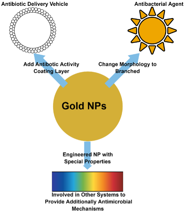
Figure 2. Schematic of gold nanoparticle engineered methods and its corresponding utilization.
2.1. Gold Nanoparticles as Antibiotic Delivery Vehicles
One of the most prominent applications of gold nanoparticles to kill bacteria include coating nanoparticles with a layer of substance with antimicrobial activity. In this case, the nanoparticle plays the role as the antibiotic delivery vehicle and would decrease the amount of antibiotic necessary to kill bacteria. [34] Technically, both chemical-synthesis and bio-synthesis methods can be used to synthesize gold nanoparticles with an antimicrobial active layer. In either synthesis method, Au3+ is reduced to Au0 and an antimicrobial substance is attached (usually adsorbed) to the surface.
Specifically, two steps are usually involved in the chemical-synthesis pathway. During the first step, Au3+ reacts with the reducing agents, such as trisodium citrate and sodium borohydride, and forms an anion capped gold nanoparticle. Within the second step, the anion surface layer in the gold nanoparticle is replaced by an antibiotic forming antibiotic capped gold nanoparticle. [35, 36] Figure 3 shows the schematic of a gold nanoparticle’s two-step chemical-synthesis pathway.
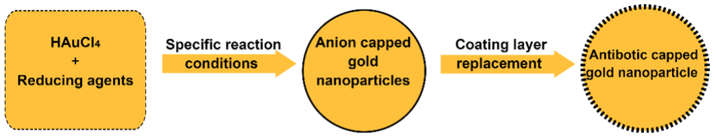
Figure 3. Two-step chemical-synthesis schematic diagram of gold nanoparticles.
For example, the citrate mediated chemical-synthesis method has been used to produce 10 nm diameter spherical gold nanoparticles coated with streptomycin, gentamicin, and neomycin surface layers. The coated nanoparticles inhibited the growth of S. aureus, M. luteus, E. coli, and P. aeruginosa. Antibiotic coated nanoparticles have better anti-bacterial performance in the Kirby-Bauer tests in general, averaging a 17.6% colony forming unit inhibition area increase than pure antibiotic at the same concentration of 0.1 mM. However, no data related to a minimum inhibitory concentration (MIC) was given within this research. [35] Moreover, the antibiotic self-mediated chemical-synthesis method has been used to produce 50 nm diameter spherical gold nanoparticles coated with amoxicillin. Amoxicillin capped gold nanoparticles inhibited the growth of E. coli after 4 hours with an MIC of 200 μg/mL. However, no data related to the antimicrobial efficiency comparison between amoxicillin capped gold nanoparticles and pure amoxicillin was provided in this research. [37] Additionally, Hayden et al. reported that single cationic-layer coated hydrophobic gold nanoparticles have the capability to react with the cell membranes of both Gram-positive and Gram-negative bacteria, and caused the aggregation of bacteria membranes, which inhibited E. coli and B. subtilis. [38] Moreover, this property was utilized to kill multi-drug-resistant (MDR) pathogenic bacteria such as MRSA, MDR P. aeruginosa, E. cloacae complex, and MDR E. coli, leading to MIC values lower than 70 nM . [39] Among those studies, only one research group discussed the cytotoxicity of nanoparticles to mammalian cells – single cationic-layer coated hydrophobic gold nanoparticles which could maintain over 80% cell viability after a long time of exposure (under the MIC). Of course, while such studies highlight promise for the use of gold nanoparticles to kill bacteria, it is clear that the field is suffering from proper experimental controls to more fully assess their efficacy.
On the other hand, bio-synthesis methods have also been used to produce gold nanoparticles. Within these pathways, both plant extracts and some specific bacteria have been used to reduce Au3+ to Au0, as well as some specific antimicrobial activity in plant cell sap or bacteria extraction substances have been coated on the surface during the process of forming gold nanoparticles. Figure 4 shows a schematic of producing gold nanoparticles via a bio-synthesis pathway.
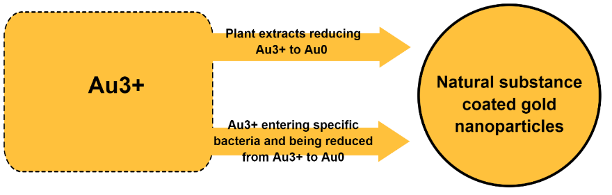
Figure 4. Bio-synthesis schematic diagram of producing gold nanoparticles.
For example, Galaxaura elongataplant and Mentha piperita extraction mediated methods were utilized to synthesize gold nanoparticles with a natural compound coated layer, and which have the capacity to kill E. coli, K. pneumoniae, and MRSA. Specifically, gold nanoparticles with a Galaxaura elongataplant extract coating layer have better antibacterial performance than the pure Galaxaura elongataplant extract, which increased an average of 57.1% colony forming inhibition area to various bacteria in Kirby-Bauer tests. [40, 41] However, no data related to MICs per specific antibacterial data related to gold nanoparticles with the Mentha piperita extract coating layer was given within the research.
Moreover, some specific bacteria such as D. radiodurans, Pseudomonas veronii AS41G, and Zooglea ramigera were used to produce gold nanoparticles, which have the capacity to inhibit S. aureus, MRSA, P. aeruginosa, group A streptococcus, M. tuberculosis, and E. coli. [42-45] However, no data related to MICs and no data related to mammalian cytotoxicity tests were given among the studies. Figure 5 provides a schematic diagram of the mechanism of the functionalized gold nanoparticles targeting and combating MDR bacteria, as adopted from reference 39.
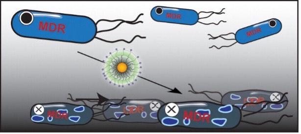
Figure 5. Schematic diagram of the mechanism of functionalized gold nanoparticles targeting and combating MDR bacteria, adopted from reference 39.
2.2. Gold Nanoparticles Alone as Anti-bacterial Agents
Unfortunately, there has been limited reported research focusing on the application of gold nanoparticles as anti-bacteria agents without an antimicrobial coating layer, yet much promise exists. It is notable that current research has found that gold nanoparticles alone have anti-bacterial properties that are dependent on their shape. [34, 46] For example, the anti-bacterial ability of branched gold nanoparticles (such nano-flowers and nano-stars) have been observed by Penders et al., in which the anti-bacterial properties of gold were shape- and size-dependent. [47] Specifically, branched gold nanoparticles with a nano-flower structure inhibited over 75% of S. aureus growth within 24 hours at a concentration of 500 μg/mL. Technically, discrete dipole approximation (DDA) simulated an enhanced electromagnetic field at the tip of the branched gold nanoparticles, which have a close correlation with its light activation property. [48, 49] Based on the localized surface plasmon resonance (LSPR) theorem, those surface electromagnetic field properties would further influence the surface electrochemical properties of a nanoparticle. [50] Biologically, the membrane potential of bacteria can affect cellular properties such as cellular proliferation and cellular respiration. [51] Thus, it is has been proven that the mechanism of the anti-bacterial properties of branched gold nanoparticles were controlled by the branched structure, or shape, of gold nanoparticles, and well-designed branched gold nanoparticles would have a strong potential for their anti-bacterial properties (without resorting to antibiotic use).
On the other hand, gold nanoparticles have a strong engineering potential to invoke complex antimicrobial systems and strengthen their bacteria inhibition by radiation sensitivity. The physical properties of gold nanoparticles (light activation, electron migration, and photothermal effect), provide additional potential to develop additional mechanisms to kill bacteria. [52, 53]
Moreover, Fasciani et al. reported an aspartame-stabilized gold-silver core-shell nanoparticle system (AuNP@Ag@Asm), which combined self-anti-bacterial capability and plasmonic photothermal property together and achieved improved antimicrobial capacity. The research reported MIC values of 3.13 µM (under a light environment, with the involvement of a photothermal mechanism) and 12.5 µM (under a dark environment, without the involvement of photothermal mechanism) to E. coli, as well MIC values of 6.25 µM (under a light environment, with the involvement of photothermal mechanism) and 12.5 µM (under a dark environment, without the involvement of photothermal mechanism) to S. aureus. [54] Additionally, Huang et al. reported a kind of polygonal shaped gold nanoparticle with a bis(vancomycin)cystamide coated layer to be used as a photothermal agent, and which has the capability to kill over eight kinds of bacteria after 5 minutes of near-infrared spectroscopy (NIR) exposure. [55] Table 1 concludes examples above morphology, synthesis method, and coating layer of AuNPs, as well as the anti-bacterial species.
Table 1. Gold Nanoparticles and Their Bacteria Inhibition Properties.
| NP Size | Morphology | Synthesis Method | Coating Layer | Bacteria | Ref |
|---|---|---|---|---|---|
| 80-100 nm | Sphere | Citrate Capped Method |
Streptomycin Gentamycin Neomycin |
S. aureus M. luteus E. coli P. aeruginosa |
35 |
| 22-52 nm | Sphere | Cefaclor Mediated | Amoxicillin | E. coli | 36, 37 |
| 100 nm | Sphere | “Bottom-up” Assembly | Hexyl-substituted, Ammonium-functionalized Thiol |
S. aureus P. aeruginosa B. subtilis E. cloacae E. coli |
38, 39 |
| 100 nm | Sphere | Galaxaura elongate Mediated | Alga Compound |
E. coli K. pneumoniae MRSA |
40 |
| 100 nm | Sphere | M. piperita Mediated | Lamiaceae Compound | E. coli | 41 |
| Unknown | Irregular |
Deinococcus radiodurans Mediated |
Unknown |
S. aureus E. coli |
42, 43 |
| 10 nm | Sphere |
Pseudomonas veronii AS41G mediated |
Unknown |
S. aureus MRSA |
44 |
| Under 20 nm | Sphere | Zooglea ramigera Mediated | Unknown |
P. aeruginosa S. aureus E. coli Group A streptococcus M. tuberculosis |
45 |
| 10 –50 nm | Nano-star | HEPES mediated | HEPES | S. aureus | 47 |
| 10-50 nm | Nanoflower | Seed mediated | Citrate acid | S. aureus | 47 |
| 10/100 nm | Nanotube | Titania Mediated Surface Modification | Unknown | Most gram-Positive and Gram-negative | 47 |
| 100 nm | Sphere | Silver Mediated Surface Modification | Aspartame | Photothermal Effect | 50 |
| 100 nm | Polygonal | Bis(vancomycin) cystamide Mediated Surface | Bis(vancomycin) cystamide |
E. coli DH5[alpha] S. aureus S. pyogenes E. coli UTI E. coli O157: H7 A. baumannii VRE MRSA PDRAB |
51 |
2.3. Mechanisms of Gold Nanoparticle Inhibition of Bacteria
Technically, there are several factors that affect gold nanoparticle bacteria inhibition. Most research has focused on the antimicrobial capability of an antimicrobial coating using gold nanoparticles as an antibiotic vehicle. In this case, gold nanoparticles were 10 nm in diameter and spherical, which was reasonable to consider since they were the first generation of gold nanoparticles developed. Thus, to date, the ability of gold nanoparticles to inhibit bacteria relied on their surface coating and in many respects could be independent of gold; that is, any nanoparticle chemistry coated with an antibiotic would kill bacteria. However, some limited studies have highlighted the importance of the gold chemistry as an antibacterial nanoparticle by highlighting the enhanced electromagnetic field that occurs at the tip of branched gold nanoparticles to inhibit S. aureus. By showing that such properties are related to the number and length of gold nano tips, it is reasonable to consider that a key property of gold nanoparticles used as antimicrobial agent is the properties of gold tip structures. [56] Additionally, another study showed a close relationship between in-plane dipole excitation and the branch structure of gold nanoparticles as confirmed with surface-enhanced Raman scattering. [57] Thus, it is reasonable to conclude that engineered gold nanoparticles have photothermal properties to combat bacteria (as they do cancer cells), and their photothermal effect is more significant in branched gold nanoparticles due to its irregular parts, such as the sharpness and length of the tips.
Moreover, engineered AuNPs have capability to involved anti-bacterial systems providing continuous electron remove ability. For example, Li et al. developed a gold-titanium-dioxide system (Au@TiO2), which coated gold onto the surface of TiO2 nanotubes by a magnetron sputtering method. The system effectively killed nearly 100% of both gram-positive and gram-negative bacteria by continuously transferring the respiratory electrons from the bacterial membrane to gold nanoparticles and eventually to TiO2. [58] Figure 6 adopted from ref. [58], provides a schematic diagram of the possible anti-bacterial mechanism of the Au@TiO2 system. Whereas in this figure Φ′ represents the work function of Au@TiO2 system, Φ represents the work function of respiratory proteins, χ represents the electron affinity of microbial membranes; EF, Ec, Ev, and EF’, Ec’, Ev’ represent the energy at Fermi level, bottom level, and top level of membrane and Au@TiO2 system, respectively.
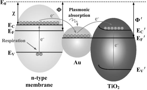
Figure 6. Schematic diagram of the possible anti-bacterial mechanism of the Au@TiO2 system, adopted from reference 58.
In conclusion, most of the current research focuses on engineering gold nanoparticles as antibiotic delivery vehicles and for additional properties involving other complicated antimicrobial systems. The most significant study could continuously transfer bacterial membrane electrons to the antimicrobial system and eventually kill both bacteria and cancer cells. Although foreseeable advantages exist in the application of gold nanoparticles as anti-bacterial agents without an antimicrobial coating layer, there is limited research focused on either the gold nanoparticles’ self-anti-bacterial capability or combined to plasmonic photothermal properties together to achieve better antimicrobial capacity. As well, although AuNPs synthesized by green chemistry have been studied in depth, the mechanism by which AuNPs selectively kill bacteria and cancer cells was not fully discussed. Thus, it is reasonable to predict that researchers should combine the gold nanoparticle’s self-anti-bacterial properties and its light activation ability, as well as the green chemically synthesized AuNPs with multi-properties for improved biological applications. In summary, again, AuNPs show promising antibacterial properties, but research has been limited in explicitly outlining, at a molecular level, why.
3. Gold Nanoparticles for Cancer Cell Inhibition
Compared to its anti-bacterial applications, gold nanoparticles have been studied in depth for numerous cancer treatments. Generally, gold nanoparticles have significant promise for cancer treatment with a combination of radiotherapy, chemotherapy, and photothermal therapy. Both mechanistic research and clinical studies proved that gold nanoparticles can enhance several cancer treatments because of their advanced physical properties. Specifically, such theoretical research simulated the light activation process of gold nanoparticles within nanoseconds, as well as the chemical process that gold nanoparticles affect cancer cell DNA activity, radical formation, and cell cycle disruption. [59] Moreover, three significant properties (the size, peak absorption wavelength, and surface area) of gold nanoparticles have all been well studied for future gold nanoparticle design.60 Based on their utilization for cancer treatment, gold nanoparticles have been successfully used as drug delivery vehicles, antibody agents, thioimidate agents, and biosensors for cancer detection. [52, 60-62] Figure 7, adopted from reference 62, highlights gold nanoparticle use and advantages for cancer treatment.
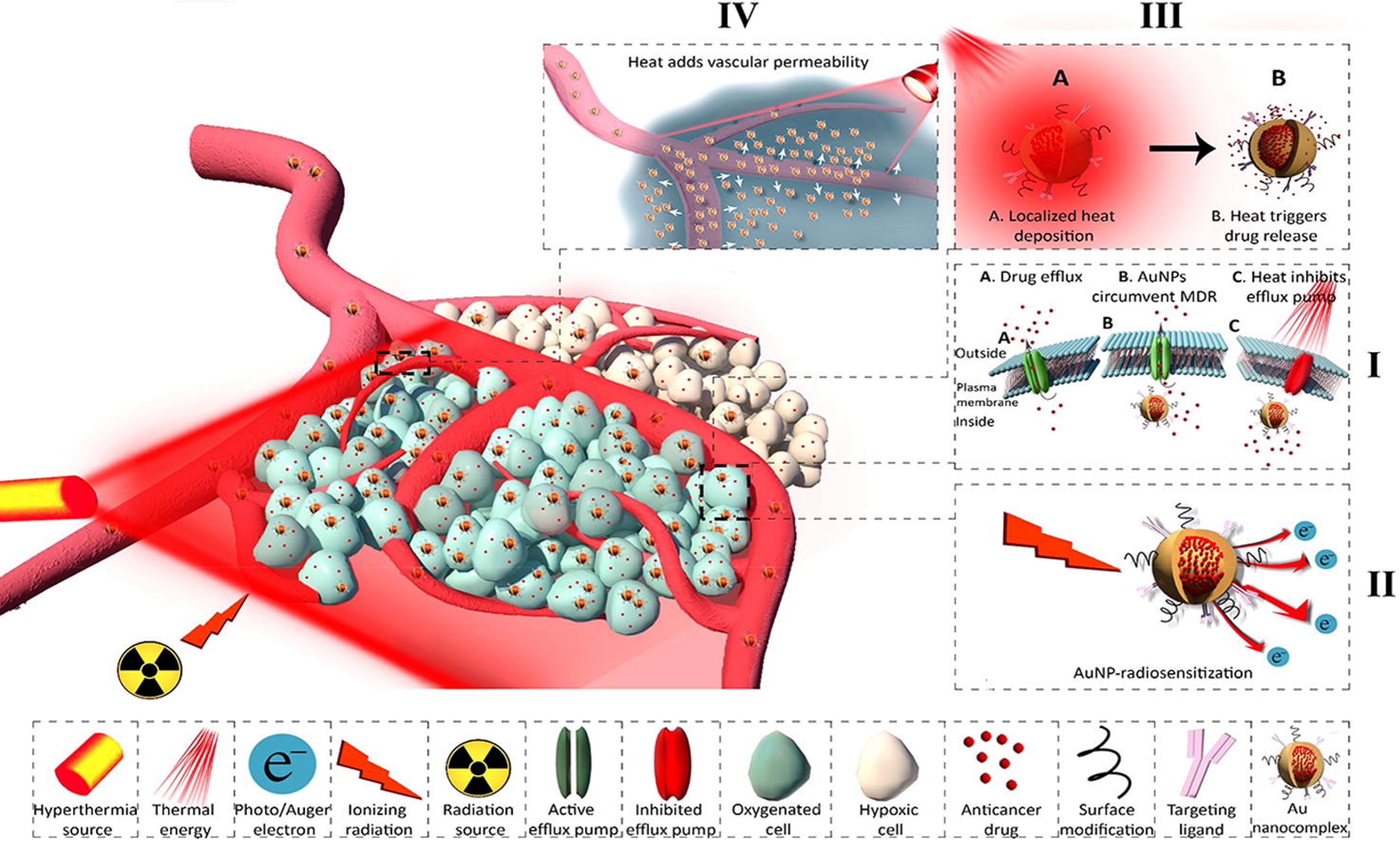
Figure 7. Usages and advantages of AuNPs in cancer treatment: (I) strategy of using AuNPs to combat MDR cancer cells; (II) strategy of using AuNPs in radiosensitization; (III) strategy of using AuNP as heat sources; and (IV) strategy of using AuNPs for increasing vascular permeability; adopted from ref. 62.
3.1. Gold Nanoparticles Used as Drug Delivery Vehicles for Cancer Treatment
Surface functionalization of gold nanoparticles is key for the cytotoxicity and anticancer properties of gold nanoparticles. [60] As discussed above for antibacterial applications, it is reasonable to consider gold nanoparticles as drug delivery vehicles for chemotherapeutic drugs as well as a functionalize gold nanoparticle surface for reacting with cancer cells. Additionally, gold nanoparticles have been widely used as both diagnostic and treatment vehicles in chemotherapy (so called “theranostic” properties). Beik et al. recently highlighted such achievements of the use of gold nanoparticles in chemotherapy. Over nine kinds of major chemotherapy drugs were successfully attached to gold nanoparticles (including doxorubicin, cisplatin, imatinib mesylate, oxaliplatin, paclitaxel, platinum, sunitinib malate, methotrexate, and bleomycin). Based on the category of the diseased organs, gold nanoparticles were used to treat numerous solid tumors, such as lung cancer, liver cancer, spleen cancer, and brain tumors. For example, bleomycin was attached to spherical gold nanoparticles with a peptide-based coating layer, which decreased the cell survival fraction of MDA-MB-231 breast cancer cells from 60% to 18% in vitro when compared to pure bleomycin at the same concentration. Imatinib mesylate was also attached to multi-layer polymer coated AuNP which decreased the proliferation of B16F10 melanoma cells from 41% to 18% when compared to pure imatinib mesylate at the same concentration. [62] However, most research focused on the molecules attached to the gold nanoparticles and the structure/geometry of the nanoparticles were not fully discussed. Furthermore, a quick review of the literature indicates much less attention being paid to the complex structure of gold nanoparticles as opposed to the drugs used.
Of course, another key area of research has been to functionalize gold nanoparticles to target the delivery of chemotherapeutics to cancer cells compared to other cells. For example, asparaginase functionalized gold joint nanoparticles have been successfully delivered to target cancer cell lines A549 and A2780 in vitro, proving their capability to treat ovarian carcinoma and lung cancer. Specifically, asparaginase functionalized AuNPs targeted and inhibited 20% of an ovarian cancer cell line A2780 at a concentration of 100 µg/ml, as well the same AuNPs targeted and inhibited over 55% of a lung cancer cell line A549 at a concentration of 125 µg/ml. Asparaginase functionalized AuNPs possessed cytotoxicity properties to cancer cells higher than individual AuNPs and asparaginase. [63]
Additionally, engineered AuNPs have been used as vehicles for cancer biosensors. For example, one study reported by Saeed et al. functionalized AuNPs with ERBB2c and CD24c and a graphene oxide underlayer (AuNPs-GO) for detecting breast cancer. Specifically, AuNP-GO possessed an excellent ability to isolate thiolated nucleic acid. Since, its sensitivity to target ERBB2c and CD24c was as low as to 378 nA/nM and 219 nA/nM, respectively, AuNP-GO can be used as an early stage breast cancer detection method.64 Thus, based on its sensitivity, AuNPs-GO increased detection efficiency, and which provides the possibility to treat the breast cancer in very early stage.
3.2. Gold Nanoparticles Used for Other Cancer Treatment Methods
Due to the sensitivity of AuNPs to radiation, they have been utilized as radiosensitizers for cancer radiotherapy. [59, 65] Furthermore, gold nanoparticles are not only involved in traditional radiotherapy but for photothermal therapy, which is a novel treatment method that can kill tumors using portion controlled high temperature since cancer cells are more sensitive to temperature increases than healthy cells. Different than traditional radiotherapy using ionizing radiation to damage lesions, photothermal therapy used specific wavelength electromagnetic radiation (such as near infrared laser and visible spectrum) which can cause portion controlled high temperature to kill cancer cells. [59, 60] In general, at least four kinds of nanostructured gold nanoparticles could be used in photothermal anti-cancer applications, which include but are not limited to nanoshells, nanorods, well designed nanospheres, and surface functionalized NIR-Tunable nanoparticles.
Specifically, gold nanoshells utilized for tumor photothermal treatment was first reported by Hirsch et al. in 2003, and magnetic resonance was required at that time for guiding the location of gold nanoparticles to tumors or cells. The research verified a significant temperature increase of gold nanoshell mediated tumor cells after 6-minutes of NIR-exposure in vivo; a maximum of 60℃ partial temperature increased in tumor cells as well as an irreversible photothermal ablation both in vitro and in vivo were observed. [66, 67] Moreover, recent research achievements include using specific porous amino-functionalized porous metal–organic framework (NH2-MOFs) coated gold nanoshells and porous gold nanoshell coated photosensitizer chlorin e6 (Ce6)-loaded nanoparticles (PUA-Ce6) as heat sources for photothermal therapy; such materials have been used in vivo and irreversible ablation was observed in a MCF-7 tumor-bearing mouse model after a 24-hour laser exposure at a power of 1.0 W/cm2 and wavelength of 808 nm. [68] Furthermore, Dong et al. reported that Fe3O4 superparamagnetic core gold shell nanoparticles effectively decreased MCF-7 tumor size in vivo with an intervention of 808 nm laser irradiation for five minutes. Fe3O4 provides superparamagnetic properties to the nanoparticles, as well as further potential applications in the magnetic resonance imaging field. [69]
Moreover, not only have gold nanoshells been involved in complex nano-designed systems and utilized for enhancing photodynamic therapy and photothermal therapy, but so have gold nanorods. For example, Sun et al. reported that several gold nanorods-capped and Ce6-doped mesoporous silica nanorods (AuNRs-Ce6-MSNRs) could be used as heat sources for photothermal and photodynamic combined therapy. In vivo experiments indicated that the AuNRs-Ce6-MSNRs group has the capability to kill over 80% of tumor cells. [70]
Not only for photothermal and photodynamic therapies, but engineered gold nanoparticles also have the capability to extenuate oxidative damage in chemotherapy as well. Specifically, Yang et al. developed a doxorubicin loaded porous Au@Pt nanoparticle modified by a cRGD peptide (DOX/Au@Pt-cRGD), which simultaneously combined the ability of increasing photothermal efficiency, enhancing chemotherapeutic effects, and mitigating chemotherapeutic effects caused by oxidative stress damage and cardiomyopathy in one nanoparticle. [71] Figure 8, adopted from reference 71, provides the synthesis process of DOX/Au@Pt-cRGD, as well the mechanism of combining these three properties into one nanoparticle.
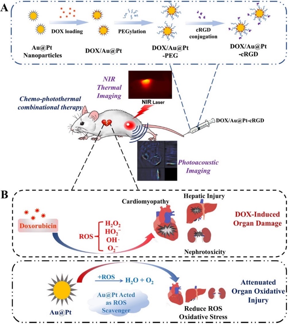
Figure 8. Mechanistic schematic of DOX/Au@Pt-cRGD combined photothermal therapy and extenuating chemotherapy damage: (A) Synthetic process of DOX/Au@Pt-cRGD and (B) Mechanism of extenuating chemotherapy damage photothermal therapy.
Similar to anti-bacterial capability, some specific biosynthesized gold nanoparticles have shown strong potential for cancer treatment. For example, Fazal et al. first reported the potential utilization of gold nanoparticles produced by a biosynthesis method in cancer treatment combined with photothermal therapy. The nanoparticles showed excellent biocompatibility with over four cell lines, and greatly inhibited an epidermoid carcinoma (A-431) cell line when exposed to NIR at wavelengths of 800 to 1000 nm. [72] Additionally, Han et al. reported E. coli inhibition using a hollow gold nanoshell complex coated silica microrod, which successfully induced T98G cell line death with the intervention of 808 nm NIR. [73] Table 2 highlights the size, morphology, utilization, coating layer, and cell type exposed to gold nanoparticles involving in cancer treatment.
Table 2. Gold Nanoparticles and Their Cancer Inhibiting Properties.
| NP Size | Morphology | Application | Coating layer | Cancer Type | Ref |
|---|---|---|---|---|---|
| Dimeter from 10 to 100 nm | Not a significant factor, normally sphere | Drug delivery vehicle in chemotherapy |
Doxorubicin, Cisplatin, Imatinib mesylate oxaliplatin, paclitaxel, Platinum, sunitinib malate, methotrexate |
Breast tumor, melanoma cells, colorectal carcinoma, prostate cancer cells |
62 |
| 35 – 55 nm | Sphere | Heat source in photothermal treatment | PEG |
Lung cancer, liver cancer, spleen cancer, brain tumor |
63 |
| 30 nm | Sphere | DNA Sensor |
ERBB2c (DNA), CD24c (DNA) |
Breast cancer | 64 |
| 120 nm | Nano-shell | Heat source in photothermal treatment | none | Human breast epithelial carcinoma SK-BR-3 cell | 66, 67 |
| 100 nm | Nano-shell | Photodynamic and photothermal synergistic therapy | Porous Au–organic frameworks | MCF-7 cell | 68 |
| 10/100 nm | Nano-rod | Photodynamic and photothermal synergistic therapy | Ce6-doped mesoporous silica | 4T1 cells | 70 |
| 100 nm | Nano-shell | Heat source in photothermal treatment | None | MCF-7 cell | 68 |
| 20 nm | Nano-sphere | Heat source in photothermal treatment | Cocoa seed extraction |
A431 cell line, MDA-MB231 cell line, L929 cell line, NIH-3T3 cell line |
72 |
| 500/2000 nm | Micro-rod | Heat source in photothermal treatment | Silica coated E. coli | T98G cell | 73 |
| 30-50 nm | Nano-star | Heat source in photothermal treatment and chemotherapy alleviating | DOX/Au@Pt-cRGD | MDA-MB-231 cells | 71 |
In conclusion, sufficient and in-depth research has proved that gold nanoparticles possess extensive applications in cancer treatment. The most current research includes irreversibly ablated AuNPs to the tumor by photothermal therapy, as well a combined three cancer treatment technique in one engineered nanoparticle. However, similar to the AuNP in the anti-bacterial field, there has been very limited research on AuNPs alone (without functionalization or coatings) directly applied to cancer cells, as well as research discussing correlations between AuNP structure and biological applications. It is reasonable to predict that future research will focus on correlations between AuNP structure property relationships and cancer treatments, as well invoking multi-properties in one AuNP (for example, antibacterial with anti-infection).
4. Mammalian Cell Cytotoxicity of Gold Nanoparticles
There are limited studies reporting on the cytotoxicity of gold nanoparticles both in vivo and in vitro. Generally, most uncoated gold nanoparticles above 10 nm in diameter have not been found to be cytotoxic to mammalian cells. For example, research has tested the cytotoxicity of 10 nm AuNPs with four different morphologies (spheres, stars, shells, and branched) using normal adult human dermal fibroblasts (HDF) with MTS cell proliferation assays and confocal microscopy. There was no significant cell proliferation inhibition observed. As well, no direct evidence was found to support the correlation between the cytotoxicity and concentration of AuNPs, and no significant toxicity was found for AuNP concentrations as high as 1 mg/mL. [47]
Some studies have indicated that the cytotoxicity of gold nanoparticles is size- and surface-functionalization-dependent. For example, an in vitro study on HeLa cervix carcinoma epithelial cells (HeLa), mouse monocytic/macrophage cells (J774A1), and mouse fibroblasts (L929) showed ignorable cytotoxicity properties of AuNPs with a diameter in 15 nm (non-significant cytotoxicity at concentration as high as to 6300 µM); however, 1-2 nm AuNPs indicated strong cell cytotoxicity with minimum IC50 values of 30 µM, 600 µM, and 56 µM to HeLa, J774A1, and L929 respectively.[74] Moreover, Goodman et al. discovered that spherical AuNPs with a cationic surface coating layer (cationic-AuNPs) pose more toxicity to Cos-1 cells and healthy human red blood cells than AuNPs with an anionic surface coating layer (anionic-AuNPs). Specifically, the LC50 value of cationic-AuNPs were 1.0 µM and 1.2 µM to Cos-1 cells and healthy human red blood cells, respectively; moreover, these values for anionic-AuNPs were higher than 7.37 µM and 72 µM, respectively. Additionally, Niidome et al. indicated that hexadecyltrimethylammonium bromide coated gold nanorods (CTAB-AuNPs) were more toxic than polyethyleneglycol coated gold nanorods (PEG-AuNPs) to Hela cells. Specifically, MTT assays indicated that CTAB-AuNPs have the capability to kill over 80% of HeLa cells after 24 hours exposure at a concentration of 0.05 mM; moreover, this value would further increase to over 95% at a concentration higher than 0.1mM. On the other hand, no significant cell toxicity was found for PEG-AuNPs, where over 90% of Hela cells survived after 24 hours of exposure at a concentration higher than 0.5 mM.[75-77]
On the other hand, within such cytotoxicity research, AuNPs have been compared to other metallic nanoparticles, with commensurate properties and in similar applications. Technically, the cytotoxicity of silver nanoparticles is closely related to the generation of reactive oxygen species (ROS). This relationship has an impact on both mammalian and bacteria cells.[47, 78, 79] Human lung fibroblast cells (IMR-90) and glioblastoma cells (U251) were utilized to test the effect of silver nanoparticles to mitochondrial damage, the generation of ROS, and potential DNA damage.[80] For example, Larese et al. indicated that sliver nanoparticles would damage HDF cells in vitro after 24 hours of exposure at a concentration of 10 ng/cm2, however, no similar damage was observed when using spherical AuNPs at the same concentration.[81] Rahman et al. used silver nanoparticles intraperitoneally injected into healthy mice, which indicated an up-regulation of metabolism and oxidative stress genes caused by silver nanoparticles potentially causing an increase in ROS and neurotoxicity.[82]
Furthermore, some studies have focused on the relationship between cellular uptake, cytotoxicity, and the mechanism of the observed cytotoxicity of gold nanoparticles. For example, Pernodet et al. focused on cell proliferation, morphological structure, and cellular activity when exposed to gold nanoparticles. The study pointed out that 14 nm diameter citrate coated spherical gold nanoparticles can enter cells by crossing the cell membrane and finally accumulate in the vacuoles of cells, although, there is no direct evidence to prove that this accumulation affects cellular function.[83] Connor et al. discovered that 18 nm spherical CTAB-capped gold nanoparticles can be taken up into K562 leukemia cells but did not show any toxicity to cells.[84] As well, Lin et al. hypothesized that the cytotoxicity of gold nanoparticles may be linked to the lipid membranes of cells and have a relationship with cellular uptake. The mechanism of the higher toxicity of cationic functionalized gold nanoparticles was related to the higher attachment of the nanoparticles to the cell membranes.[85]
Critically, less ROS generation has been found for gold nanoparticles than silver nanoparticles. For example, as reported by Li et al., less ROS production from the gold nanoparticles was measured intracellularly than silver nanoparticles within a D. radiodurans protein extract mediated system.[43] Although the conditions of the experiments were limited to a protein extract mediated system, since less oxidation of gold nano particles existed over silver nanoparticles, it is reasonable to predict that less ROS production would occur in vivo. It is notable that the cytotoxicity of silver nanoparticles is relevant to its oxidative behavior, which can cause inflammatory, genotoxic, and the potential DNA damage.[86] In summary, the size, shape, and surface properties could affect cytotoxicity. Critical cytotoxicity was always accompanied by properties of small size and toxic coating layers. Based on the fact that AuNPs used in cancer treatment and anti-bacterial applications were 10 -100 nm in dimeter range and coated with non-toxic coating layers, the AuNPs recommended above would have very limited toxicity to human cells.
5. Conclusions and Future Prospects
In conclusion, this article reviewed gold nanoparticles used to treat bacterial infection, cancer treatment, and their mammalian cell cytotoxicity properties. Studies have uniformly demonstrated that gold nanoparticles exhibit extreme promise in the antimicrobial field and may be used as novel antibiotic delivery vehicles, anti-bacterial agents, and become involved in complex anti-bacterial systems providing additional antimicrobial mechanisms. The morphology and activity are different and vary according to the gold nanoparticles in terms of functionalization, size, and shape. Specifically, the activity of an antibiotic layer and branched structure, respectively, are critical when gold nanoparticles are used as an antibiotic delivery vehicle and anti-bacterial agent. When gold nanoparticles are involved in a complex anti-bacterial system, its position is normally as the heat source after the activation by specific IR wavelengths. Similarly, for many decades, gold nanoparticles promise extensive anti-cancer properties. Its utilization includes but is not limited to serving as a drug delivery vehicle for chemotherapeutic agents, a heat source in photothermal treatment, an enhancer for photodynamic and photothermal synergistic therapy, etc. The morphology of gold nanoparticles involved in cancer treatments is normally an engineered nano-shell, nanosphere, and nano-rod. AuNPs combined multi-properties into one nanoparticle (such as the DOX/Au@Pt-cRGD and Au@TiO2 system) would gain significant achievement in the foreseeable future.
On the other hand, gold nanoparticles have demonstrated excellent biocompatibility in terms of limited mammalian cell cytotoxicity, surpassing that of other nanoparticle chemistries. Except for 1 nm diameter gold nanospheres, nearly all other nanoparticles with various morphologies (sphere, star, and nano-shell) have not been found to be significantly toxic to mammalian cells. Intracellular accumulation has been observed, however, no relationship between accumulation and cell function has been reported. Furthermore, cytotoxicity comparison experiments indicate less oxidative behavior and genotoxic of gold than silver nanoparticles. Thus, it is reasonable to predict that gold nanoparticles have more utilization in biological applications than other metallic nanoparticles. It is notable that although applications of gold nanoparticle in the anti-bacterial and anti-cancer fields have been well studied, limited systemic studies have been found. Such research is critical due to the close relationship between bacteria and cancer and well as the increased susceptibility of infection among cancer patients. Future research should focus on the mechanism of the inhibition effect and reaction with mammalian cells. Future studies should also determine the genetic responses from cells, possible gold nanoparticle resistivity in bacteria and cancer cells (similar to what has been developed for antibiotics and chemotherapeutic agents), and in vivo confirmation of promising in vitro results; all of which are imperative for this field to progress.
The COVID-19 pandemic has imparted both transformative shifts and promising opportunities to this research field. On one hand, the pandemic disrupted a multitude of fundamental research endeavors. It is noteworthy that contemporary fundamental research continues to focus on mechanisms consistent with those explored five years ago, wherein the integration of photothermal therapy and drug delivery remains a predominant research theme.[87] On the other hand, emerging technologies such as RNA-based strategies and targeted therapeutic approaches have furnished novel insights for researchers. NPs can function as efficient delivery vectors, while the functional characteristics of specific AuNPs necessitate further in-depth investigation.[88] The convergence of AuNPs with machine learning is exceedingly scarce—a research gap that merits heightened attention, given that advanced therapeutic technologies are increasingly contingent upon big data as a foundational database.
Furthermore, translational research serves a pivotal role in facilitating the translation of these laboratory-developed technologies into practical clinical applications. Beyond bridging the "bench-to-bedside" gap, translational studies have become increasingly critical in response to the pandemic-driven demand for rapid, scalable, and clinically viable solutions. This includes validating the safety and efficacy of NP-based drug delivery systems in preclinical and clinical trials, optimizing manufacturing processes to ensure cost-effectiveness and mass production, and addressing regulatory hurdles associated with novel therapeutic platforms. Notably, the pandemic has accelerated interdisciplinary collaboration between material scientists, pharmacologists, clinicians, and regulatory experts, fostering a more streamlined translational pipeline.[89] Additionally, translational efforts have focused on adapting NP-mediated delivery for antiviral agents (e.g., remdesivir-loaded polymeric NPs) to improve bioavailability and tissue penetration, addressing unmet clinical needs in treating severe viral infections.[90, 91] Real-world data generated from these translational initiatives has further provided valuable feedback for refining fundamental research directions—such as optimizing NP surface modifications to reduce immunogenicity based on clinical trial observations—creating a synergistic loop that enhances both the scientific rigor and practical relevance of the field.
Acknowledgement
The authors gratefully acknowledge the guidance provided by Prof. Dr. Thomas Jay Webster.
Funding
No funding.
Disclosure
The authors have nothing to disclose.
References
-
“World health statistics 2018: monitoring health for the SDGs sustainable development goals.” 2018.
-
C. Årdal, E. Baraldi, F. Ciabuschi, K. Outterson, J. H. Rex, L. J. Piddock, D. Findlay. “To the G20: incentivising antibacterial research and development.” Lancet Infect. Dis. 2017, 17, 8, 799–801.
-
E. Pursey, D. Sünderhauf, W. H. Gaze, E. R. Westra, S. van Houte. “CRISPR-cas antimicrobials: challenges and future prospects.” PLOS Pathog. 2018, 14, 6, e1006990.
-
D. I. Andersson, D. Hughes. “Persistence of antibiotic resistance in bacterial populations.” FEMS Microbiol. Rev. 2011, 35, 5, 901–11.
-
T. J. Webster, I. Seil. “Antimicrobial applications of nanotechnology: methods and literature.” Int. J. Nanomed. 2012, 2767.
-
F. L. Nobrega, A. R. Costa, L. D. Kluskens, J. Azeredo. "Revisiting phage therapy: new applications for old resources." Trends Microbiol. 2015, 23, 4, 185–91.
-
Y. K. Kang, K. Kwon, J. S. Ryu, H. N. Lee, C. Park, H. J. Chung. "Nonviral genome editing based on a polymer-derivatized CRISPR nanocomplex for targeting bacterial pathogens and antibiotic resistance." Bioconjugate Chem. 2017, 28, 4, 957–67.
-
E. C. Keen. “Phage therapy: concept to cure.” Front. Microbiol. 2012, 3, 238.
-
“Cancer trends progress report 2018.” NATIONAL CANCER INSTITUTE at the National Institutes of Health, 2018.
-
P. Sharma, S. Hu-Lieskovan, J. A. Wargo, A. Ribas. “Primary, adaptive, and acquired resistance to cancer immunotherapy.” Cell 2017, 168, 4, 707–23.
-
A. Titov, A. Petukhov, A. Staliarova, D. Motorin, E. Bulatov, O. Shuvalov, S. M. Soond, M. Piacentini, G. Melino, A. Zaritskey. “The biological basis and clinical symptoms of CAR-T therapy-associated toxicites.” Cell Death Dis. 2018, 9, 9, 897.
-
D. Hanahan, R. A. Weinberg. “Hallmarks of cancer: the next generation.” Cell 2011, 144, 5, 646–74.
-
L. A. Doyle, W. Yang, L. V. Abruzzo, T. Krogmann, Y. Gao, A. K. Rishi, D. D. Ross. “A multidrug resistance transporter from human MCF-7 breast cancer cells.” Proc. Natl. Acad. Sci. 1998, 95, 26, 15665–70.
-
A. K. Iyer, A. Singh, S. Ganta, M. M. Amiji. “Role of integrated cancer nanomedicine in overcoming drug resistance.” Adv. Drug Delivery Rev. 2013, 65, 13–14, 1784–802.
-
S. Wu, S. Powers, W. Zhu, Y. A. Hannun. “Substantial contribution of extrinsic risk factors to cancer development.” Nature 2016, 529, 7584, 43–47.
-
J. Parsonnet. “Bacterial infection as a cause of cancer.” Environ. Health Perspect. 1995, 103, Suppl 8, 263–68.
-
J. T. Schiller, D. R. Lowy. “Virus infection and human cancer: an overview.” 2014, 193, 1–10.
-
D. Van Elsland, J. Neefjes. “Bacterial infections and cancer.” EMBO Rep. 2018, 19, 11, e46632.
-
IARC Working Group on the Evaluation of Carcinogenic Risks to Humans. "Biological agents." IARC Monogr. Eval. Carcinog. Risks Hum. 2012, 100, Pt B, 1–441.
-
T. Scanu, R. M. Spaapen, J. M. Bakker, C. B. Pratap, L. Wu, I. Hofland, A. Broeks, V. K. Shukla, M. Kumar, H. Janssen. "Salmonella manipulation of host signaling pathways provokes cellular transformation associated with gallbladder carcinoma." Cell Host Microbe 2015, 17, 6, 763–74.
-
L. Mughini-Gras, M. Schaapveld, J. Kramers, S. Mooij, E. A. Neefjes-Borst, W. van Pelt, J. Neefjes. "Increased colon cancer risk after severe salmonella infection." PLOS One 2018, 13, 1, e0189721.
-
H. Zhu, Z. Shen, H. Luo, W. Zhang, X. Zhu. "Chlamydia trachomatis infection-associated risk of cervical cancer: a meta-analysis." Medicine (Baltimore) 2016, 95, 13, e3077.
-
B. Trabert, T. Waterboer, A. Idahl, N. Brenner, L. A. Brinton, J. Butt, S. B. Coburn, P. Hartge, K. Hufnagel, F. Inturrisi. "Antibodies against chlamydia trachomatis and ovarian cancer risk in two independent populations." JNCI: J. Natl. Cancer Inst. 2019, 111, 2, 129–36.
-
S. Ascioglu, J. H. Rex, B. De Pauw, J. E. Bennett, J. Bille, F. Crokaert, D. W. Denning, J. P. Donnelly, J. E. Edwards, Z. Erjavec. "Defining opportunistic invasive fungal infections in immunocompromised patients with cancer and hematopoietic stem cell transplants: an international consensus." Проблемы Медицинской Микологии 2003, 5, 1, 10–16.
-
G. P. Bodey, M. Mardani, H. A. Hanna, M. Boktour, J. Abbas, E. Girgawy, R. Y. Hachem, D. P. Kontoyiannis, I. I. Raad. "The epidemiology of candida glabrata and candida albicans fungemia in immunocompromised patients with cancer." Am. J. Med. 2002, 112, 5, 380–85.
-
S. Ray, R. Mohan, J. K. Singh, M. K. Samantaray, M. M. Shaikh, D. Panda, P. Ghosh. "Anticancer and antimicrobial metallopharmaceutical agents based on palladium, gold, and silver N-heterocyclic carbene complexes." J. Am. Chem. Soc. 2007, 129, 48, 15042–53.
-
C. D. Brown, D. M. Cruz, A. K. Roy, T. J. Webster. "Synthesis and characterization of PVP-coated tellurium nanorods and their antibacterial and anticancer properties." J. Nanopart. Res. 2018, 20, 9, 254.
-
H. Zeng, G. F. Combs Jr. "Selenium as an anticancer nutrient: roles in cell proliferation and tumor cell invasion." J. Nutr. Biochem. 2008, 19, 1, 1–7.
-
D. Medina Cruz, G. Mi, T. J. Webster. "Synthesis and characterization of biogenic selenium nanoparticles with antimicrobial properties made by staphylococcus aureus, methicillin‐resistant staphylococcus aureus (MRSA), escherichia coli, and pseudomonas aeruginosa." J. Biomed. Mater. Res., Part A 2018, 106, 5, 1400–12.
-
R. A. Sperling, P. R. Gil, F. Zhang, M. Zanella, W. J. Parak. "Biological applications of gold nanoparticles." Chem. Soc. Rev. 2008, 37, 9, 1896–908.
-
X. Zhang. "Gold nanoparticles: recent advances in the biomedical applications." Cell Biochem. Biophys. 2015, 72, 3, 771–75.
-
A. Khan. "Medicine at nanoscale: a new horizon." Int. J. Nanomed. 2012, 2997.
-
V. V. Kumar, S. P. Anthony. "Antimicrobial studies of metal and metal oxide nanoparticles." 2016, 265–300.
-
Y. Zhang, T. P. Shareena Dasari, H. Deng, H. Yu. "Antimicrobial activity of gold nanoparticles and ionic gold." J. Environ. Sci. Health C 2015, 33, 3, 286–327.
-
A. N. Grace, K. Pandian. "Antibacterial efficacy of aminoglycosidic antibiotics protected gold nanoparticles—a brief study." Colloids Surf., A 2007, 297, 1–3, 63–70.
-
A. Rai, A. Prabhune, C. C. Perry. "Antibiotic mediated synthesis of gold nanoparticles with potent antimicrobial activity and their application in antimicrobial coatings." J. Mater. Chem. 2010, 20, 32, 6789–98.
-
M. Demurtas, C. C. Perry. "Facile one-pot synthesis of amoxicillin-coated gold nanoparticles and their antimicrobial activity." Gold Bull. 2014, 47, 1–2, 103–07.
-
S. C. Hayden, G. Zhao, K. Saha, R. L. Phillips, X. Li, O. R. Miranda, V. M. Rotello, M. A. El-Sayed, I. Schmidt-Krey, U. H. F. Bunz. "Aggregation and interaction of cationic nanoparticles on bacterial surfaces." J. Am. Chem. Soc. 2012, 134, 16, 6920–23.
-
X. Li, S. M. Robinson, A. Gupta, K. Saha, Z. Jiang, D. F. Moyano, A. Sahar, M. A. Riley, V. M. Rotello. "Functional gold nanoparticles as potent antimicrobial agents against multi-drug-resistant bacteria." ACS Nano 2014, 8, 10, 10682–86.
-
N. Abdel-Raouf, N. M. Al-Enazi, I. B. Ibraheem. "Green biosynthesis of gold nanoparticles using galaxaura elongata and characterization of their antibacterial activity." Arabian J. Chem. 2017, 10, S3029–39.
-
D. MubarakAli, N. Thajuddin, K. Jeganathan, M. Gunasekaran. "Plant extract mediated synthesis of silver and gold nanoparticles and its antibacterial activity against clinically isolated pathogens." Colloids Surf., B 2011, 85, 2, 360–65.
-
J. Li, Q. Li, X. Ma, B. Tian, T. Li, J. Yu, S. Dai, Y. Weng, Y. Hua. "Biosynthesis of gold nanoparticles by the extreme bacterium deinococcus radiodurans and an evaluation of their antibacterial properties." Int. J. Nanomed. 2016, 11, 5931–44.
-
J. Li, B. Tian, T. Li, S. Dai, Y. Weng, J. Lu, X. Xu, Y. Jin, R. Pang, Y. Hua. "Biosynthesis of Au, Ag and Au–Ag bimetallic nanoparticles using protein extracts of deinococcus radiodurans and evaluation of their cytotoxicity." Int. J. Nanomed. 2018, 13, 1411–24.
-
S. Baker, S. Satish. "Biosynthesis of gold nanoparticles by pseudomonas veronii AS41G inhabiting annona squamosa L." Spectrochim. Acta, Part A 2015, 150, 691–95.
-
N. Srivastava, M. Mukhopadhyay. "Biosynthesis and characterization of gold nanoparticles using zooglea ramigera and assessment of its antibacterial property." J. Cluster Sci. 2015, 26, 3, 675–92.
-
R. M. Amin, M. B. Mohamed, M. A. Ramadan, T. Verwanger, B. Krammer. "Rapid and sensitive microplate assay for screening the effect of silver and gold nanoparticles on bacteria." Nanomed. 2009, 4, 6, 637–43.
-
J. Penders, M. Stolzoff, D. J. Hickey, M. Andersson, T. J. Webster. "Shape-dependent antibacterial effects of non-cytotoxic gold nanoparticles." Int. J. Nanomed. 2017, 12, 2457–68.
-
W.-H. Yang, G. C. Schatz, R. P. Van Duyne. "Discrete dipole approximation for calculating extinction and raman intensities for small particles with arbitrary shapes." J. Chem. Phys. 1995, 103, 3, 869–75.
-
K. L. Kelly, E. Coronado, L. L. Zhao, G. C. Schatz. "The optical properties of metal nanoparticles: the influence of size, shape, and dielectric environment." J. Phys. Chem. B 2003, 107, 3, 668–77.
-
C. Wang, X.-G. Nie, Y. Shi, Y. Zhou, J.-J. Xu, X.-H. Xia, H.-Y. Chen. "Direct plasmon-accelerated electrochemical reaction on gold nanoparticles." ACS Nano 2017, 11, 6, 5897–905.
-
J. P. Stratford, C. L. A. Edwards, M. J. Ghanshyam, D. Malyshev, M. A. Delise, Y. Hayashi, M. Asally. "Electrically induced bacterial membrane-potential dynamics correspond to cellular proliferation capacity." Proc. Natl. Acad. Sci. 2019, 116, 19, 9552–57.
-
L. C. Kennedy, L. R. Bickford, N. A. Lewinski, A. J. Coughlin, Y. Hu, E. S. Day, J. L. West, R. A. Drezek. "A new era for cancer treatment: gold‐nanoparticle‐mediated thermal therapies." Small 2011, 7, 2, 169–83.
-
A. M. Alkilany, C. J. Murphy. "Toxicity and cellular uptake of gold nanoparticles: what we have learned so far?" J. Nanopart. Res. 2010, 12, 7, 2313–33.
-
C. Fasciani, M. J. Silvero, M. A. Anghel, G. A. Argüello, M. C. Becerra, J. C. Scaiano. "Aspartame-stabilized gold–silver bimetallic biocompatible nanostructures with plasmonic photothermal properties, antibacterial activity, and long-term stability." J. Am. Chem. Soc. 2014, 136, 50, 17394–97.
-
W.-C. Huang, P.-J. Tsai, Y.-C. Chen. "Functional gold nanoparticles as photothermal agents for selective-killing of pathogenic bacteria." Nanomed. 2007, 2, 6, 777–87.
-
E. Hao, R. C. Bailey, G. C. Schatz, J. T. Hupp, S. Li. "Synthesis and optical properties of 'branched' gold nanocrystals." Nano Lett. 2004, 4, 2, 327–30.
-
O. M. Bakr, B. H. Wunsch, F. Stellacci. "High-yield synthesis of multi-branched urchin-like gold nanoparticles." Chem. Mater. 2006, 18, 14, 3297–301.
-
J. Li, H. Zhou, S. Qian, Z. Liu, J. Feng, P. Jin, X. Liu. "Plasmonic gold nanoparticles modified titania nanotubes for antibacterial application." Appl. Phys. Lett. 2014, 104, 26.
-
S. Her, D. A. Jaffray, C. Allen. "Gold nanoparticles for applications in cancer radiotherapy: mechanisms and recent advancements." Adv. Drug Delivery Rev. 2017, 109, 84–101.
-
D. A. Giljohann, D. S. Seferos, W. L. Daniel, M. D. Massich, P. C. Patel, C. A. Mirkin. "Gold nanoparticles for biology and medicine." Spherical Nucleic Acids 2020, 55–90.
-
M. Z. Ahmad, S. Akhter, Z. Rahman, S. Akhter, M. Anwar, N. Mallik, F. J. Ahmad. "Nanometric gold in cancer nanotechnology: current status and future prospect." J. Pharm. Pharmacol. 2013, 65, 5, 634–51.
-
J. Beik, M. Khateri, Z. Khosravi, S. K. Kamrava, S. Kooranifar, H. Ghaznavi, A. Shakeri-Zadeh. "Gold nanoparticles in combinatorial cancer therapy strategies." Coord. Chem. Rev. 2019, 387, 299–324.
-
G. Baskar, B. G. Garrick, K. Lalitha, M. Chamundeeswari. "Gold nanoparticle mediated delivery of fungal asparaginase against cancer cells." J. Drug Delivery Sci. Technol. 2018, 44, 498–504.
-
A. A. Saeed, J. L. A. Sánchez, C. K. O’Sullivan, M. N. Abbas. "DNA biosensors based on gold nanoparticles-modified graphene oxide for the detection of breast cancer biomarkers for early diagnosis." Bioelectrochemistry 2017, 118, 91–99.
-
A. M. Gobin, E. M. Watkins, E. Quevedo, V. L. Colvin, J. L. West. "Near‐infrared‐resonant gold/gold sulfide nanoparticles as a photothermal cancer therapeutic agent." Small 2010, 6, 6, 745–52.
-
S. J. Oldenburg, R. D. Averitt, S. L. Westcott, N. J. Halas. "Nanoengineering of optical resonances." Chem. Phys. Lett. 1998, 288, 2–4, 243–47.
-
L. R. Hirsch, R. J. Stafford, J. A. Bankson, S. R. Sershen, B. Rivera, R. E. Price, J. D. Hazle, N. J. Halas, J. L. West. "Nanoshell-mediated near-infrared thermal therapy of tumors under magnetic resonance guidance." Proc. Natl. Acad. Sci. 2003, 100, 23, 13549–54.
-
C. Liu, L. Luo, L. Zeng, J. Xing, Y. Xia, S. Sun, L. Zhang, Z. Yu, J. Yao, Z. Yu, O. U. Akakuru, M. Saeed, A. Wu. "Porous gold nanoshells on functional NH2‐MOFs: facile synthesis and designable platforms for cancer multiple therapy." Small 2018, 14, 35, 1801851.
-
W. Dong, Y. Li, D. Niu, Z. Ma, J. Gu, Y. Chen, W. Zhao, X. Liu, C. Liu, J. Shi. "Facile synthesis of monodisperse superparamagnetic Fe3O4 core@hybrid@Au shell nanocomposite for bimodal imaging and photothermal therapy." Adv. Mater. 2011, 23, 45, 5392–97.
-
Q. Sun, Q. You, X. Pang, X. Tan, J. Wang, L. Liu, F. Guo, F. Tan, N. Li. "A photoresponsive and rod-shape nanocarrier: single wavelength of light triggered photothermal and photodynamic therapy based on AuNRs-capped & Ce6-doped mesoporous silica nanorods." Biomaterials 2017, 122, 188–200.
-
Q. Yang, J. Peng, Y. Xiao, W. Li, L. Tan, X. Xu, Z. Qian. "Porous Au@Pt nanoparticles: therapeutic platform for tumor chemo-photothermal co-therapy and alleviating doxorubicin-induced oxidative damage." ACS Appl. Mater. Interfaces 2018, 10, 1, 150–64.
-
S. Fazal, A. Jayasree, S. Sasidharan, M. Koyakutty, S. V. Nair, D. Menon. "Green synthesis of anisotropic gold nanoparticles for photothermal therapy of cancer." ACS Appl. Mater. Interfaces 2014, 6, 11, 8080–89.
-
S. Han, Y.-J. Park, E.-J. Park, Y. Kim. "T98G cell death induced by photothermal treatment with hollow gold nanoshell-coupled silica microrods prepared from escherichia coli." ACS Appl. Mater. Interfaces 2019, 11, 9, 8831–37.
-
Y. Pan, S. Neuss, A. Leifert, M. Fischler, F. Wen, U. Simon, G. Schmid, W. Brandau, W. Jahnen‐Dechent. "Size‐dependent cytotoxicity of gold nanoparticles." Small 2007, 3, 11, 1941–49.
-
C. J. Murphy, A. M. Gole, J. W. Stone, P. N. Sisco, A. M. Alkilany, E. C. Goldsmith, S. C. Baxter. "Gold nanoparticles in biology: beyond toxicity to cellular imaging." Acc. Chem. Res. 2008, 41, 12, 1721–30.
-
C. M. Goodman, C. D. McCusker, T. Yilmaz, V. M. Rotello. "Toxicity of gold nanoparticles functionalized with cationic and anionic side chains." Bioconjugate Chem. 2004, 15, 4, 897–900.
-
T. Niidome, M. Yamagata, Y. Okamoto, Y. Akiyama, H. Takahashi, T. Kawano, Y. Katayama, Y. Niidome. "PEG-modified gold nanorods with a stealth character for in vivo applications." J. Controlled Release 2006, 114, 3, 343–47.
-
H. J. Johnston, G. Hutchison, F. M. Christensen, S. Peters, S. Hankin, V. Stone. "A review of the in vivo and in vitro toxicity of silver and gold particulates: particle attributes and biological mechanisms responsible for the observed toxicity." Crit. Rev. Toxicol. 2010, 40, 4, 328–46.
-
C. Marambio-Jones, E. M. V. Hoek. "A review of the antibacterial effects of silver nanomaterials and potential implications for human health and the environment." J. Nanopart. Res. 2010, 12, 5, 1531–51.
-
P. V. AshaRani, G. Low Kah Mun, M. P. Hande, S. Valiyaveettil. "Cytotoxicity and genotoxicity of silver nanoparticles in human cells." ACS Nano 2009, 3, 2, 279–90.
-
F. F. Larese, F. D’Agostin, M. Crosera, G. Adami, N. Renzi, M. Bovenzi, G. Maina. "Human skin penetration of silver nanoparticles through intact and damaged skin." Toxicology 2009, 255, 1–2, 33–37.
-
M. F. Rahman, J. Wang, T. A. Patterson, U. T. Saini, B. L. Robinson, G. D. Newport, R. C. Murdock, J. J. Schlager, S. M. Hussain, S. F. Ali. "Expression of genes related to oxidative stress in the mouse brain after exposure to silver-25 nanoparticles." Toxicol. Lett. 2009, 187, 1, 15–21.
-
N. Pernodet, X. Fang, Y. Sun, A. Bakhtina, A. Ramakrishnan, J. Sokolov, A. Ulman, M. Rafailovich. "Adverse effects of citrate/gold nanoparticles on human dermal fibroblasts." Small 2006, 2, 6, 766–73.
-
E. E. Connor, J. Mwamuka, A. Gole, C. J. Murphy, M. D. Wyatt. "Gold nanoparticles are taken up by human cells but do not cause acute cytotoxicity." Small 2005, 1, 3, 325–27.
-
J. Lin, H. Zhang, Z. Chen, Y. Zheng. "Penetration of lipid membranes by gold nanoparticles: insights into cellular uptake, cytotoxicity, and their relationship." ACS Nano 2010, 4, 9, 5421–29.
-
S. Arora, J. Jain, J. M. Rajwade, K. M. Paknikar. "Cellular responses induced by silver nanoparticles: in vitro studies." Toxicol. Lett. 2008, 179, 2, 93–100.
-
P. Singh, S. Pandit, S. R. Balusamy, M. Madhusudanan, H. Singh, H. M. Amsath Haseef, I. Mijakovic. "Advanced nanomaterials for cancer therapy: gold, silver, and iron oxide nanoparticles in oncological applications." Adv. Healthcare Mater. 2025, 14, 4, 2403059.
-
L. Zhuang, Y. Lian, T. Zhu. "Multifunctional gold nanoparticles: bridging detection, diagnosis, and targeted therapy in cancer." Mol. Cancer 2025, 24, 1, 228.
-
E. Kole, K. Jadhav, R. Singh, R. K. Verma, A. Chatterjee, J. Naik. "Next-generation nanoparticle-enabled mRNA vaccines in the treatment of COVID-19." Crit. Rev. Ther. Drug Carr. Syst. 2025, 42, 6, 83–123.
-
M. Zhou, S. Xiang, Y. Zhao, Y. Tang, J. Yang, X. Yin, J. Tian, S. Hu, Y. Du. "[68Ga]Ga-AUNP-12 PET imaging to assess the PD-L1 status in preclinical and first-in-human study." Eur. J. Nucl. Med. Mol. Imaging 2024, 51, 2, 369–79.
-
S. Bevers, S. A. Kooijmans, E. Van de Velde, M. J. Evers, S. Seghers, J. J. Gitz-Francois, N. C. van Kronenburg, M. H. Fens, E. Mastrobattista, L. Hassler. "mRNA-LNP vaccines tuned for systemic immunization induce strong antitumor immunity by engaging splenic immune cells." Mol. Ther. 2022, 30, 9, 3078–94.