A Mini Review of Nano-Structured Metal for Cancer Treatment
published: 18 July 2025 | https://doi.org/10.63174/xdi.ZFGQ3834
Abstract
Nanostructured metal materials have attracted much attention in cancer treatment field because of their special properties, like large surface area, tunable shape and size, and good physical and chemical stability. Many kinds of nanostructured metal can be used in different cancer therapy methods, such as photothermal therapy, radiotherapy, Reactive oxygen species (ROS) generation, and drug delivery. With the development of controllable synthesis methods, progressively morphologies such as rod, shell, cage, and branched type can be made, which benefit to strong response in light, magnetic field, or radial-frequency (RF), and applied in specific targeted lesion area. These metal nanomaterials also can be used for combined treatment and diagnosis. In this mini review, we introduce recent progress of nanostructured metal in cancer therapy, and also talk about their working mechanism, structure design, and possible problems for future clinical applications.
1. Introduction
Cancer remains a leading threat to human health worldwide, with recent statistics estimating nearly 20 million new cases and 10 million deaths globally in 2020 alone, and these numbers are expected to rise steadily in the coming decades.[1] Although advancements in conventional treatment options—such as chemotherapy, radiotherapy, and immunotherapy—have improved patient outcomes in many cases, persistent obstacles remain. These include treatment resistance, toxicity to healthy tissues, and limited effectiveness in advanced or heterogeneous tumors.[2] The need for more targeted, efficient, and less invasive treatment strategies has become increasingly urgent.
In response to these challenges, researchers have begun to explore nanostructured metals as innovative therapeutic platforms in oncology. These materials, engineered at the nanoscale, offer distinct physical and chemical properties that allow precise interactions with cancer cells and tumor environments. Unlike traditional drugs that directly interact with cancer cells, nano-structured metal serves multiple role in treatment, it includes acting as direct cytotoxic agents, enabling controlled drug release, or mediating therapies activated by light, heat, or magnetic fields.[3–6] Metals such as silver, platinum, copper, iron oxide, and tellurium have each demonstrated potential in various therapeutic contexts, including photothermal therapy (PTT), photodynamic therapy (PDT), chemodynamical therapy (CDT), and magnetic hyperthermia.[7–9]
The diversity of nanoparticle geometries—ranging from spheres and rods to more complex branched or hollow structures—enables tuning of optical absorption, cellular uptake, and biodistribution profiles.[10–13] These features support the development of highly localized therapies that spare surrounding healthy tissue, an essential requirement for clinical translation. Moreover, several of these metal nanomaterials are being integrated into diagnostic–therapeutic systems, allowing simultaneous imaging and treatment of tumors.
Despite promising laboratory and animal model results, major hurdles remain. These include incomplete understanding of long-term toxicity, behavior in complex biological systems, and challenges related to reproducible large-scale synthesis. Continued interdisciplinary research will be necessary to optimize nanostructure design, improve safety, and enable successful clinical implementation.
This mini review, cataloged by the morphology of nanostructured metals, outlines the recent progress in cancer therapy application, covering their mechanisms of action, structural design principles, and the key challenges that must be addressed to move toward real-world applications.
2. Application of Nanostructured Metals Cataloged by Morphology
2.1. Spherical Nanoparticles
Spherical nanostructured metal particles are among the most widely applied forms in cancer therapy due to their isotropic geometry, scalable synthesis, and favorable surface modification properties. Their uniform morphology and controllable size distribution enables predictable interactions with biological systems, making them ideal candidates for applications such as ROS generation, photothermal therapy (PTT), photodynamic therapy (PDT), and radiosensitization.[14,15]
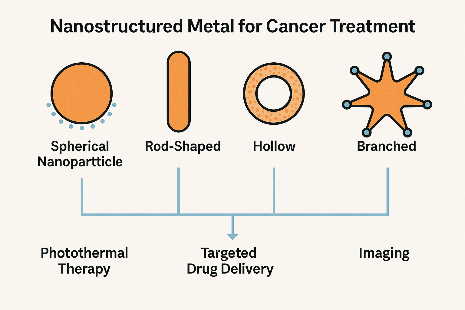
Figure 1. oncept illustration of how nanostructured metal is used in cancer treatments
One of the primary uses of spherical metal nanoparticles is in photothermal therapy, where localized heating is generated upon irradiation with near-infrared (NIR) or visible light. Unlike conventional radiotherapy, which utilizes ionizing radiation to damage DNA, PTT relies on the thermal conversion properties of nanoparticles to induce localized heat sufficient to ablate cancerous tissue. Spherical particles made from silver, iron oxide, platinum, copper, and other metals can be tuned for optimal light absorption and photothermal efficiency. Surface coatings such as polyethylene glycol (PEG), silica, or organic ligands are often applied to improve biocompatibility and tumor targeting.[16–18]
In recent years, multi-functional spherical nanoparticles have been developed for combined therapy approaches. For example, iron oxide–platinum core–shell spheres have been engineered to deliver simultaneous photothermal therapy and MRI-based tumor imaging. Similarly, porous or polymer-coated spherical nanoparticles have been used to load chemotherapeutic agents or photosensitizers for synergistic photothermal–chemotherapy or photothermal–PDT applications.[19]
Furthermore, biosynthesized spherical nanoparticles—prepared using microbial or plant extracts—have demonstrated promising cancer treatment results due to their biocompatibility and natural surface functionalities.[20] These biologically derived particles have shown photothermal effects under NIR irradiation and effective tumor cell inhibition across various cancer types, including breast cancer, glioblastoma, and epidermoid carcinoma.
Spherical nanostructures are also extensively used as drug carriers. Their geometry allows for moderate drug-loading capacity in a few years ago and surface functionalization, which can be leveraged for controlled release in response to external stimuli (e.g., pH, temperature, or light).[21] Common chemotherapeutics such as doxorubicin, cisplatin, paclitaxel, and methotrexate have been incorporated into spherical metal nanoparticles, enhancing both tumor accumulation via the enhanced permeability and retention (EPR) effect and therapeutic efficacy.
Overall, spherical nanostructured metals open up a new era of utilizing nanoparticles as a tool to treat cancers, providing a highly versatile platform in cancer nanomedicine, combining therapeutic, diagnostic, and delivery capabilities in a single, tunable system.
2.2. Rod-Shaped and Wire-Like Nanostructures
Rod-shaped and wire-like nanostructured metals have emerged as versatile platforms in cancer therapy due to their anisotropic geometry, which allows for tunable optical, magnetic, and surface properties. Compared with spherical nanoparticles, these elongated structures often exhibit enhanced photothermal conversion efficiency, deeper tissue penetration under near-infrared (NIR) irradiation, and improved cellular uptake due to their high aspect ratio.[22,23]
One of the primary advantages of nanorods and nanowires is their shape-dependent localized surface plasmon resonance (LSPR).[24] Metal nanorods such as those composed of silver, copper, and platinum can be tuned to absorb strongly in the NIR range, making them ideal candidates for photothermal therapy. These particles convert incident NIR light into localized heat, enabling the thermal ablation of tumor tissues with minimal harm to surrounding healthy cells. Their elongated shape also improves their interaction with cell membranes, enhancing endocytosis and intracellular accumulation.
Several studies have reported successful use of iron oxide nanowires or Fe₃O₄ nanorods for magnetic hyperthermia, in which alternating magnetic fields induce localized heating to destroy cancer cells.[25] These materials can also be magnetically guided to tumor regions, offering site-specific treatment with minimal off-target effects. In combination with radiotherapy or chemotherapy, magnetic nanorods can further enhance treatment outcomes.
Rod-shaped nanoparticles also serve as multifunctional platforms for drug loading and photodynamic activation. For example, platinum nanorods or Pt–Au hybrid rods have been incorporated into drug delivery systems that release payloads under photothermal stimulation, while also catalyzing the production of reactive oxygen species (ROS) for synergistic photodynamic effects.[26,27] Furthermore, nanowires composed of copper-based compounds have demonstrated both cytotoxic and catalytic activity, disrupting the tumor microenvironment while serving as drug carriers.
Recently, biomimetic nanorods and mesoporous nanowire composites have been engineered with surface functionalization strategies—such as peptide ligands, antibodies, or PEG coatings—to enhance tumor selectivity and reduce systemic toxicity.[28,29] These elongated structures offer large surface areas for modification and can carry therapeutic agents either by adsorption or encapsulation.
In addition to their therapeutic capabilities, rod- and wire-like nanostructures are also attractive for diagnostic imaging. Due to their high metal content and anisotropic geometry, they provide enhanced contrast in photoacoustic imaging, computed tomography (CT), and magnetic resonance imaging (MRI). This enables real-time monitoring of therapeutic progress, a key advantage for clinical translation.
Overall, rod-shaped and wire-like nanostructures offer promising avenues for precise and multimodal cancer therapy. Their shape-driven functionalities—ranging from enhanced photothermal effects to guided targeting—make them critical components in the next generation of metal-based nanomedicines.
2.3. Hollow and Cage-like Structures
Hollow and cage-like nanostructures have attracted considerable attention in cancer therapy due to their high surface-to-volume ratio, internal cavity for drug loading, tunable wall thickness, and stimuli-responsive release behavior.[30] These structures provide significant advantages over solid nanoparticles, particularly in enabling controlled cargo delivery, multimodal treatment, and reduced density for improved circulation and tissue penetration.
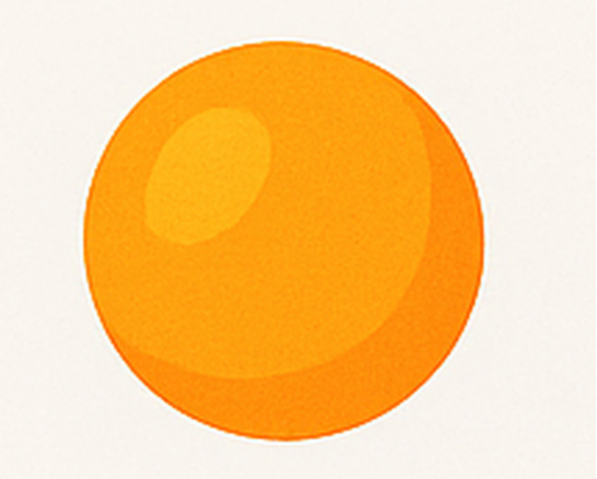
Figure 2. Concept illustration of spherical nanostructured metal (nanoparticle)
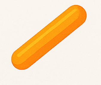
Figure 3. Concept illustration of rod-shape nanostructured metal (nanoparticle)
Figure 4. Concept illustration of hollow nanostructured metal (nanoparticle)
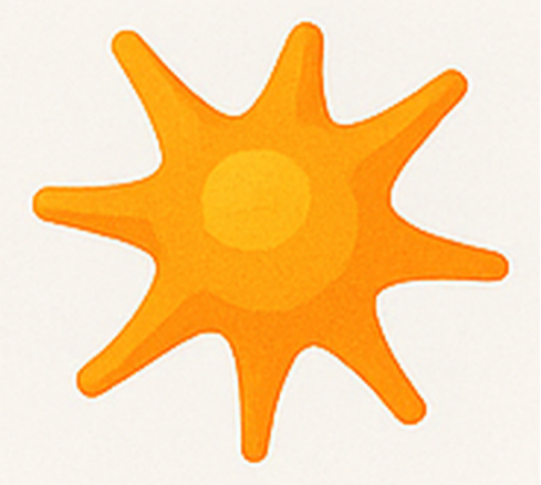
Figure 5. Concept illustration of branched nanostructured metal (nanoparticle)
Metal-based hollow nanoparticles—including those made from gold, silver, copper sulfide, and iron oxide—have been widely studied for photothermal therapy (PTT).[31] Their hollow architecture enables efficient NIR light absorption and heat generation, while also allowing interior loading of chemotherapeutic drugs, photosensitizers, or imaging agents. This architecture supports synergistic therapy strategies, combining PTT with chemotherapy or photodynamic therapy (PDT).[32–34]
One classical example is the use of metal nanoshells, such as hollow gold nanoshells, where a dielectric core (often silica) is coated with a thin metallic layer. This design provides strong plasmonic resonance in the NIR region and has been successfully employed for both imaging and hyperthermic tumor ablation. In recent developments, these nanoshells have been further modified with porous metal-organic framework (MOF) coatings, pH-responsive polymers, or targeting ligands to improve selectivity and payload release.[35]
Cage-like structures, such as mesoporous metal frameworks, allow even higher drug loading capacity. Their open architecture facilitates diffusion-controlled release, which can be externally triggered by pH, redox environment, or laser irradiation. For instance, CuS nanocages and Pt-based hollow cages have been developed for effective chemodynamical therapy (CDT) and ROS-based tumor killing in acidic tumor microenvironments.[36]
Recent reports also include biomimetic hollow nanostructures, such as silica-coated hollow gold nanospheres templated by bacteria or microrods, which show promise for deep-tissue NIR photothermal therapy and immunogenic cell death induction. These structures not only deliver therapeutic agents but may also serve as immune adjuvants, amplifying anti-tumor immune responses.
Furthermore, some hollow metal nanostructures are inherently multifunctional, offering platforms for diagnosis and therapy in one system (theranostics). For example, hollow Fe₃O₄-based nanostructures have been used for magnetic resonance imaging (MRI) and magnetic hyperthermia, while simultaneously delivering chemotherapy drugs and generating heat under alternating magnetic fields.[37]
In summary, hollow and cage-like metal nanostructures offer an advanced design strategy for multi-agent delivery, controlled release, and multi-modal therapy, making them a highly versatile toolset in the landscape of cancer nanomedicine.
2.4. Branched and Star-Shaped Nanostructures
Branched and star-shaped nanostructures are distinguished by their highly irregular surface topographies, which provide significantly enhanced surface area, multiple “hot spots” for localized electromagnetic field enhancement, and unique optical properties due to multipolar plasmon resonance modes. These features make them especially attractive for photothermal therapy (PTT), photodynamic therapy (PDT), and multifunctional theranostics in cancer treatment.[38,39]
Unlike spherical or rod-shaped particles, branched nanostructures can support strong near-infrared (NIR) absorption even at relatively small sizes. This enables efficient light-to-heat conversion at lower laser intensities, reducing thermal damage to healthy tissues. For example, silver and gold nanostars, as well as platinum-coated branched nanoparticles, have shown enhanced photothermal responses and improved tumor ablation under NIR laser irradiation.[40,41]
In addition to their photothermal properties, branched structures provide multivalent binding sites, which are advantageous for drug loading, targeting ligand attachment, and biomolecular sensing. The extended and highly curved branches increase the density of active surface regions, allowing simultaneous incorporation of multiple therapeutic agents or imaging probes. For example, DOX-loaded Au@Pt-cRGD nanostars have been designed to deliver chemotherapy drugs while simultaneously enhancing PTT and reducing systemic toxicity.[42,43]
Recent studies also demonstrate that branched metal nanostructures can improve reactive oxygen species (ROS) generation when used as catalysts in photodynamic therapy or chemodynamic therapy. For instance, branched copper oxide or iron oxide nanostars can catalyze Fenton or Fenton-like reactions in the acidic tumor microenvironment, triggering oxidative stress and promoting tumor cell apoptosis.[44]
Moreover, the sharp tips of star-shaped particles act as plasmonic “hot spots,” enhancing scattering and absorption cross-sections. This property is highly beneficial for photoacoustic imaging, surface-enhanced Raman scattering (SERS), and computed tomography (CT) contrast, enabling their use in diagnostic–therapeutic (theranostic) platforms.[45]
Surface engineering is often applied to branched nanostructures to improve colloidal stability, biocompatibility, and targeting specificity. Functionalization with polyethylene glycol (PEG), targeting peptides (e.g., RGD), or pH-responsive polymers allows these nanomaterials to selectively accumulate in tumors and release therapeutic payloads in response to the tumor microenvironment.[46]
Overall, branched and star-shaped metal nanostructures offer superior photothermal efficiency, multimodal loading capacity, and optical tunability, making them powerful candidates for integrated cancer treatment strategies combining therapy and diagnosis
3. Conclusion
Nanostructured metal-based materials offer a powerful and versatile platform for advancing cancer therapy. By tailoring their physical morphology—whether as spheres, rods, cages, branches, or core–shell structures—researchers have developed a wide range of nanomaterials capable of addressing critical challenges in cancer treatment, including poor selectivity, drug resistance, and systemic toxicity.
Each structural category presents distinct advantages: spherical nanoparticles provide uniform distribution and drug-loading efficiency; rod-shaped and wire-like structures enable deeper tissue penetration and enhanced cellular uptake; hollow and cage-like designs offer high loading capacity and controlled release; branched nanostructures maximize photothermal conversion and surface functionalization; and core–shell systems support multimodal imaging and combination therapy through architectural modularity.
These engineered nanostructures have demonstrated broad applicability across multiple therapeutic modes, including photothermal therapy (PTT), photodynamic therapy (PDT), chemodynamical therapy (CDT), magnetic hyperthermia, and targeted drug delivery. Many have also been integrated with diagnostic elements, enabling real-time monitoring and theragnostic precision.
Despite promising preclinical results, the clinical translation of these nanostructures remains limited by challenges in large-scale synthesis, long-term safety, and in vivo behavior. Continued interdisciplinary efforts in material design, surface engineering, and biological evaluation will be essential to unlock their full potential.
As cancer therapies shift toward more personalized and minimally invasive approaches, nanostructured metals—particularly when designed with structural precision and multifunctionality—are poised to play a significant role in the next generation of cancer treatment strategies.
Consent for publication
Not applicable.
Competing interests
The authors declare no competing interests.
Funding
This work is kindly supported by Shandong Provincial Natural Science Foundation (ZR2024QB360). We also gratefully acknowledge Shandong Agriculture and Engineering University Start-Up Fund for Talented Scholars (2024GCCZR-01, BSQJ202301).
Author Contribution
This study is grateful to all the authors for their full contributions.
References
H. Sung, J. Ferlay, R. L. Siegel, M. Laversanne, I. Soerjomataram, A. Jemal, F. Bray. "Global Cancer Statistics 2020: GLOBOCAN Estimates of Incidence and Mortality Worldwide for 36 Cancers in 185 Countries." CA A Cancer J Clinicians 2021, 71, 3, 209–49.
R. L. Siegel, K. D. Miller, N. S. Wagle, A. Jemal. "Cancer statistics, 2023." CA. Cancer J. Clin. 2023, 73, 1.
H. Tiwari, N. Rai, S. Singh, P. Gupta, A. Verma, A. K. Singh, Kajal, P. Salvi, S. K. Singh, V. Gautam. "Recent advances in nanomaterials-based targeted drug delivery for preclinical cancer diagnosis and therapeutics." B.Asel. 2023, 10, 7, 760.
P. Pashootan, F. Saadati, H. Fahimi, M. Rahmati, R. Strippoli, A. Zarrabi, M. Cordani, M. A. Moosavi. "Metal-based nanoparticles in cancer therapy: exploring photodynamic therapy and its interplay with regulated cell death pathways." Int. J. Pharm. 2024, 649, 123622.
S. M. Hosseini, J. Mohammadnejad, R. Najafi-Taher, Z. B. Zadeh, M. Tanhaei, S. Ramakrishna. "Multifunctional Carbon-Based Nanoparticles: Theranostic Applications in Cancer Therapy and Diagnosis." ACS Appl. Bio Mater. 2023, 6, 4, 1323–38.
P. Manivasagan, A. Joe, H.-W. Han, T. Thambi, M. Selvaraj, K. Chidambaram, J. Kim, E.-S. Jang. "Recent advances in multifunctional nanomaterials for photothermal-enhanced Fenton-based chemodynamic tumor therapy." Mater. Today Bio. 2022, 13, 100197.
M. Overchuk, R. A. Weersink, B. C. Wilson, G. Zheng. "Photodynamic and Photothermal Therapies: Synergy Opportunities for Nanomedicine." ACS Nano 2023, 17, 9, 7979–8003.
D. Aebisher, K. Rogóż, A. Myśliwiec, K. Dynarowicz, D. Bartusik-Aebisher. "The use of photodynamic therapy in medical practice." Front. Oncol. 2024, 14, 1373263.
X. Wang, X. Zhong, Z. Liu, L. Cheng. "Recent progress of chemodynamic therapy-induced combination cancer therapy." Nano Today. 2020, 35, 100946.
Y. Zhao, Y. Wang, F. Ran, Y. Cui, C. Liu, Q. Zhao, Y. Gao, D. Wang, S. Wang. "A comparison between sphere and rod nanoparticles regarding their in vivo biological behavior and pharmacokinetics." Sci. Rep. 2017, 7, 1, 4131.
N. D. Donahue, H. Acar, S. Wilhelm. "Concepts of nanoparticle cellular uptake, intracellular trafficking, and kinetics in nanomedicine." Adv. Drug Deliv. Rev. 2019, 143, 68–96.
G. D. Kalaycioglu, B. O. Altas, N. Aydogan. "Biophysical interaction of gold nanoparticles with model biological membranes: Investigation of the effect of geometry on cellular uptake and photothermal performance." Colloids Surf. Physicochem. Eng. Asp. 2023, 676, 132221.
M. J. Mitchell, M. M. Billingsley, R. M. Haley, M. E. Wechsler, N. A. Peppas, R. Langer. "Engineering precision nanoparticles for drug delivery." Nat. Rev. Drug Discov. 2021, 20, 2, 101–24.
Z. Li, Y. Chen, Y. Yang, Y. Yu, Y. Zhang, D. Zhu, X. Yu, X. Ouyang, Z. Xie, Y. Zhao. "Recent advances in nanomaterials-based chemo-photothermal combination therapy for improving cancer treatment." Front. Bioeng. Biotechnol. 2019, 7, 293.
S. M. Hosseini, J. Mohammadnejad, R. Najafi-Taher, Z. B. Zadeh, M. Tanhaei, S. Ramakrishna. "Multifunctional Carbon-Based Nanoparticles: Theranostic Applications in Cancer Therapy and Diagnosis." ACS Appl. Bio Mater. 2023, 6, 4, 1323–38.
W. He, G. Ma, Q. Shen, Z. Tang. "Engineering gold nanostructures for cancer treatment: spherical nanoparticles, nanorods, and atomically precise nanoclusters." Nanomaterials. 2022, 12, 10, 1738.
L. S. Mbatha, J. Akinyelu, C. I. Chukwuma, M. P. Mokoena, T. Kudanga. "Current trends and prospects for application of green synthesized metal nanoparticles in cancer and COVID-19 therapies." Viruses. 2023, 15, 3, 741.
J. T. Cole, N. B. Holland. "Multifunctional nanoparticles for use in theranostic applications." Drug Deliv. and Transl. Res. 2015, 5, 3, 295–309.
A. Maximenko, J. Depciuch, N. Łopuszyńska, M. Stec, Ż. Światkowska-Warkocka, V. Bayev, P. M. Zieliński, J. Baran, J. Fedotova, W. P. Węglarz. "Fe3O4@SiO2@Au nanoparticles for MRI-guided chemo/NIR photothermal therapy of cancer cells." RSC Adv. 2020, 10, 44, 26508–20.
J. B. Vines, J.-H. Yoon, N.-E. Ryu, D.-J. Lim, H. Park. "Gold nanoparticles for photothermal cancer therapy." Biomedicines. 2019, 7, 167.
L. C. Kennedy, L. R. Bickford, N. A. Lewinski, A. J. Coughlin, Y. Hu, E. S. Day, J. L. West, R. A. Drezek. "A New Era for Cancer Treatment: Gold‐Nanoparticle‐Mediated Thermal Therapies." Small. 2011, 7, 2, 169–83.
H. S. Han, K. Y. Choi. "Advances in nanomaterial-mediated photothermal cancer therapies: toward clinical applications." Biomedicines. 2021, 9, 3, 305.
Y. Zhang, Y. Zhang, G. Zhang, J. Wu, L. Wang, Z. Dong, Y. Zheng, Q. Huang, M. Zou, R. Liao. "Recent advances and clinical challenges of phototherapeutic nanoparticles in cancer monotherapy or combination therapy." Coord. Chem. Rev. 2024, 518, 216069.
X. Zhong, X. Dai, Y. Wang, H. Wang, H. Qian, X. Wang. "Copper‐based nanomaterials for cancer theranostics." WIREs Nanomed Nanobiotechnol 2022, 14, 4, e1797.
P. Q. Thong, L. T. Thu Huong, N. D. Tu, H. T. My Nhung, L. Khanh, D. H. Manh, P. H. Nam, N. X. Phuc, J. Alonso, J. Qiao, S. Sridhar, H. P. Thu, M. H. Phan, N. T. Kim Thanh. "Multifunctional Nanocarriers of Fe3O4@PLA-PEG/Curcumin for MRI, Magnetic Hyperthermia and Drug Delivery." Nanomedicine (Lond.) 2022, 17, 22, 1677–93.
H. A. Alqahtani, D. M. Alshangiti, N. Aldaleeli, M. Madani, S. A. Al‐Gahtany, M. M. Ghobashy. "Synergistic Gold Nanorod‐based Chemo‐Photothermal Therapy: A Promising Nanoparticle Approach for Refractory Multidrug‐Resistant Cancer." ChemBioEng Reviews 2024, 11, 4, e202300046.
M. Aioub, S. R. Panikkanvalappil, M. A. El-Sayed. "Platinum-Coated Gold Nanorods: Efficient Reactive Oxygen Scavengers That Prevent Oxidative Damage toward Healthy, Untreated Cells during Plasmonic Photothermal Therapy." ACS Nano. 2017, 11, 1, 579–86.
J. Zhao, H. Chen, Y. Tang, H. Chen, G. Chen, Y. Yin, G. Li. "Research progresses on the functional polypeptides in the detection and imaging of breast cancer." J. Mater. Chem. 2018, 6, 17, 2510–23.
T. Niidome, M. Yamagata, Y. Okamoto, Y. Akiyama, H. Takahashi, T. Kawano, Y. Katayama, Y. Niidome. "PEG-modified gold nanorods with a stealth character for in vivo applications." J. Controlled Release. 2006, 114, 3, 343–47.
J. Huang, Z. Xu, Y. Jiang, W. Law, B. Dong, X. Zeng, M. Ma, G. Xu, J. Zou, C. Yang. "Metal organic framework-coated gold nanrod as an on-demand drug delivery platform for chemo-photothermal cancer therapy." J Nanobiotechnol 2021, 19, 1, 219.
L. Li, L. H. Rashidi, M. Yao, L. Ma, L. Chen, J. Zhang, Y. Zhang, W. Chen. "CuS nanoagents for photodynamic and photothermal therapies: Phenomena and possible mechanisms." Photodiagnosis Photodyn. Ther. 2017, 19, 5–14.
C. Bastiancich, A. Da Silva, M.-A. Estève. "Photothermal therapy for the treatment of glioblastoma: potential and preclinical challenges." Front. Oncol. 2021, 10, 610356.
Y. Li, J. Yang, X. Sun. "Reactive oxygen species-based nanomaterials for cancer therapy." Front. Chem. 2021, 9, 650587.
E. Yasun, S. Gandhi, S. Choudhury, R. Mohammadinejad, F. Benyettou, N. Gozubenli, H. Arami. "Hollow micro and nanostructures for therapeutic and imaging applications." J. Drug Deliv. Sci. Technol. 2020, 60, 102094.
J. Kong, M. Cai, R. Zhu, Y. Zhang, Y. Du, X. Jing, Y. Sun, R. Chang, C. Qu, X. Dong. "The utilization of metal-organic frameworks in tumor-targeted drug delivery systems." J. Sci. Adv. Mater. Devices. 2024, 9, 3, 100770.
J. Zhao, L. Liu, L. Gu, Z. Li, Y. Li, Z. Wu, B. Sun, X. Wang, T. Sun. "Dual-responsive metal organic framework for electrically-enhanced cascade catalytic tumor therapy." Mater. Today Adv. 2023, 17, 100329.
H. Bunzen, D. Jirák. "Recent Advances in Metal–Organic Frameworks for Applications in Magnetic Resonance Imaging." ACS Appl. Mater. Interfaces. 2022, 14, 45, 50445–62.
T. Vo-Dinh, Y. Liu, B. M. Crawford, H.-N. Wang, H. Yuan, J. K. Register, C. G. Khoury. "Shining gold nanostars: From cancer diagnostics to photothermal treatment and immunotherapy." J. Immunol. Sci. 2018, 2, 1, 1.
J. B. Vines, J.-H. Yoon, N.-E. Ryu, D.-J. Lim, H. Park. "Gold nanoparticles for photothermal cancer therapy." Front. Chem. 2019, 7, 167.
F. Xia, J. Niu, Y. Hong, C. Li, W. Cao, L. Wang, W. Hou, Y. Liu, D. Cui. "Matrix metallopeptidase 2 targeted delivery of gold nanostars decorated with IR-780 iodide for dual-modal imaging and enhanced photothermal/photodynamic therapy." Acta Biomater. 2019, 89, 289–99.
A. L. Siegel, G. A. Baker. "Bespoke nanostars: synthetic strategies, tactics, and uses of tailored branched gold nanoparticles." Nanoscale Adv.2 2021, 3, 14, 3980–4004.
F. Zhang, L. Xu, Y. Wang, P. Wang. "Engineering Plasmonic Au Nanostars: Enhanced Plasmonic Properties, Preparation and Biomedical Application." Mater. Today Bio. 2025, 33, 102022.
X. He, S. Chen, X. Mao. "Utilization of metal or non-metal-based functional materials as efficient composites in cancer therapies." RSC advances. 2022, 12, 11, 6540–51.
R. Lu, Y. Zheng, M. Wang, J. Lin, Z. Zhao, L. Chen, J. Zhang, X. Liu, L. Yin, Y. Chen. "Reactive oxygen species-responsive branched poly (β-amino ester) with robust efficiency for cytosolic protein delivery." Acta Biomater. 2022, 152, 355–66.
B. Diloknawarit, E. N. Schaumann, T. W. Odom. "High-Curvature Features on Branched Nanoconstructs Circumvent Protein Corona Interference." ACS Appl. Mater. Interfaces 2025, 17, 10, 15187–95.