Research on Structural Design and Mechanical Properties of Biodegradable Cerebrovascular Stents
published: 31 July 2025 | https://doi.org/10.63174/xdi.FCEC3246
Abstract
This study presents a systematic investigation into the structural design and mechanical performance optimization of next-generation biodegradable stents specifically engineered for cerebrovascular applications. By addressing critical clinical demands in the treatment of cerebrovascular diseases, the research highlights the limitations of conventional cardiovascular stents, particularly their insufficient flexibility and suboptimal vascular adaptability in delicate cerebral vessels. Through comparative analysis of various structural design methodologies—including modular, non-modular, and hybrid configurations—the study identifies key performance determinants, with a focused examination of support unit geometry and connection unit arrangement. The findings demonstrate the successful development of an innovative stent architecture that integrates axially aligned sinusoidal connection units with strategically distributed discrete support elements. This novel design achieves a remarkable balance between enhanced flexibility and maintained radial strength, overcoming a major trade-off in conventional stent engineering. Furthermore, the research establishes quantitative correlations between critical structural parameters (e.g., strut width, thickness, and unit spacing) and performance metrics such as bending compliance and radial support capability, providing a robust theoretical framework for optimized stent design. To validate these advancements, the study introduces a comprehensive finite element analysis (FEA)-based evaluation methodology, which can simulate the stress and deformation of the stent under physiological conditions such as vascular bending and blood pulsation, enabling precise assessment of stent performance under simulated physiological conditions. These insights not only advance the field of biodegradable stent technology but also offer practical technical guidance for future research and development, such as prioritizing the optimization of the curvature radius of the connecting unit (initial value suggested to be 0.5–1 mm) to enhance stent flexibility. Ultimately, this work contributes significantly to the evolution of specialized stent systems tailored for the unique biomechanical challenges of cerebrovascular disease intervention.
1. Introduction
Hemorrhagic stroke has an extremely high rate of disability and mortality, with cerebral aneurysm being a representative condition[1]. Due to the sudden onset of the disease, 10% to 15% of aneurysm patients die suddenly before they can seek medical attention, and the case-fatality rate for the first bleeding is 35%, while the rate for recurrent bleeding is 60% to 80%. Even if patients survive, most are left with disabilities[2]. Endovascular interventional therapy has become the primary treatment for cerebral aneurysms due to its advantages of minimal invasiveness, fewer sequelae, broad applicability, and rapid recovery[3-7]. Stent-assisted coil embolization is a common approach in endovascular interventional therapy, effectively treating various types of aneurysms, significantly improving the embolization rate of cerebral aneurysms, and reducing their recurrence rate. However, the success of this treatment relies on the vascular stent being able to reach the target location smoothly and function safely.
Most of the current stent research focuses on treating cardiovascular diseases[8, 9], and their structural design and mechanical properties are unsuitable for treating cerebrovascular diseases. The main reason is that the cerebrovascular anatomy is more complex than the cardiovascular system[10]. Compared with cardiovascular vessels, cerebrovascular vessels have smaller diameters (usually 2–5 mm vs. 5–15 mm for cardiovascular vessels), thinner walls (approximately 0.1–0.3 mm vs. 0.3–0.8 mm for cardiovascular vessels), and more tortuous physiological curves, such as the S-shaped curve of the internal carotid artery siphon segment. During endovascular procedures, stents are typically inserted through a puncture site in the inguinal region and delivered through the abdominal aorta, thoracic aorta, and carotid artery until they reach the vicinity of the cerebral Willis circle[11]. Therefore, the clinical use of most existing stent structures is limited in the treatment of cerebrovascular diseases, necessitating further optimization of stent design and mechanical performance evaluation tailored to the anatomical characteristics of cerebral vessels. This article provides an overview of the development, structural design, and key mechanical properties of biodegradable stents, which are an emerging hotspot in the medical field, specifically in the context of cerebrovascular applications.
2. Development of Biodegradable Cerebrovascular Stents
The original intention of the vascular stent is to treat cardiovascular lumen stenosis or occlusion, and to ensure blood circulation through higher radial support. With the development of medical standards, the application of stents by medical personnel is no longer limited to cardiovascular applications in the large lumen. In 1991, an Italian scholar, Guglielmi[12], published a report on the use of vascular stents to treat cerebrovascular diseases opened up a new application space for it. In 1997, scholar Higashida[13] first tried to use a balloon-diffusion cardiovascular stent-assisted spring coil to treat cerebral artery stenosis. However, due to the unique anatomical structure in the skull, the small diameter of the blood vessels, the tortuous tube diameter, the thin and brittle blood vessel walls, which leads to poor passage of the cardiovascular system, and the damage to the blood vessels during the implantation process, ultimately leading to higher complications[1]. To meet clinical needs, cerebrovascular stents that focus on flexibility came into being[14-17]. Currently, commercial cerebrovascular stents used for stent-assisted embolization treatment include Neuroform stents from Stacyk in the United States[18], Enterprise stents from Johnson & Johnson in the United States[19], Leo stents from Bulgge in France[20], and Apollo stents from China[21], etc., where (a) the Neuroform stent features an open-grid structure and (d) the Apollo stent has a closed-loop design. No matter which commercial cerebrovascular stents are, these are permanent implants due to material reasons. If a permanent stent is implanted in the blood vessel, it will always exist in the form of a foreign body, continuously stimulating the blood vessel wall, causing new problems, such as the blood vessels being unable to restore their original physiological functions, advanced stent thrombosis, advanced vascular restenosis, mismatch between the implanted site and the stent-free site, endothelial dysfunction, etc. And for brain aneurysms with high recurrence rates, such stents will directly affect their secondary intervention[22, 23].
With the introduction of the concept of "degradable", the concept of biodegradable cardiovascular and cerebrovascular stents has emerged. At present, biodegradable cardiovascular stents have been successfully used in clinical treatment[24-26], but biodegradable cerebrovascular stents are still in the early stages of development, and no clinical reports have been seen. By comparison, the relatively slow progress in the development of biodegradable cerebrovascular stents is primarily attributable to the unique anatomical characteristics of cerebral vasculature, which impose significant constraints on the clinical application of current vascular stent technologies. The anatomy of the cerebrovascular system is extremely complex, and some parts are extremely tortuous, such as the siphon section of the internal carotid artery, which can form relatively sharp angles in a narrow space. Therefore, considering the unique anatomical characteristics of cerebrovascular and the flexibility of blood vessels after stent deployment, the existing vascular stent structure still has the risk of not reaching the target and serving safely[1]. In addition, according to performance requirements, the design idea of cerebrovascular stents is different from that of cardiovascular stents. The structure of the cardiovascular stent focuses on its support, and the primary factor in the development of a new cerebrovascular stent should be its flexibility[1]. Good flexibility ensures that the stent and its delivery system can reach the target position smoothly, and ensures that the stent is perfectly fitted with the complex arterial wall after release.
This paper provides a detailed analysis of the structural design problems of insufficient mechanical properties of existing cerebral vascular stents and combines the latest research results.
3. Research on the Structural Design of Biodegradable Cerebrovascular Stents
At present, there are no complete and unified design standards and specifications for vascular stents at home and abroad, and the skeleton structure design of the stent is completely based on experience. The design prototype of the stent skeleton structure determines whether the subsequent optimization can be carried out based on experimental feedback to quickly obtain the final results that meet clinical needs. Understanding and mastering the current design of different stent structures and their correspondence with mechanical properties is conducive to the design of the new cerebrovascular stent structure prototype. There are many studies on the design of stent structure, and they will be reviewed in three parts according to their research purpose.
3.1. Stent Scaffold Structure
The existing vascular stent structures are mostly designed based on a modular stent structure design method. The components of such stent structures are defined separately in the ASTM F2681-06 standard[27, 28]: support unit and bridge reinforcement. The support unit, which can represent the individual element of the smallest unit of the radial support part of the stent, is represented as a circular cross-section in the circumferential direction and radial direction of the stent. The bridge rib, also known as the connecting unit, is the connecting part between the radial support units of the stent. Compared with the support unit, the bridge ribs can have unique design characteristics to improve the mechanical properties of the stent. The annular support unit combined with the discrete connection unit is the standard frame that most support structure designs currently follow. Wei et al.[29] designed an unequal high-supporting unit structure through the design scheme of increasing the angle of the support unit after expansion. The radial supportability of this type of stent increased by 40% compared with the Abbott BVS stent structure. Dun[30] designed an axial symmetry stent. The annular support unit of the stent is relatively complex, but it significantly improves the expansion uniformity of the stent. Wang et al.[31] designed a zero Poisson's ratio stent. The support unit of the Z-ring structure solves the axial shortening problem during coronary stent expansion. Song et al.[32] designed a two-strut stent based on the structure of the commercial stent BVS. The simulation results show that the stent has better radial stiffness and axial shortening compared to the BVS stent. Shobayashi et al.[33] designed a close-cell stent. The flexibility of the stent is only slightly lower than that of the Neuroform stent. Due to its close-cell stent design, the "crocodile back" phenomenon of the open-loop stent was successfully avoided during the bending process[34]—also known as the "fish scale" phenomenon[35]. However, this study also indirectly shows that the modular stent design method may have certain limitations in improving flexibility.
Some researchers have tried to design the scaffold using other design methods, and its mechanical properties have also been improved accordingly. Xia et al.[36] designed a chiral constrained stent and knitted stent based on node-line poles. The axis shortening rate and dog-boning rate when the scaffold is expanded are almost zero, the radial resilience is small, and the plaque prolapse rate is low. Singh et al.[37] designed a bionic scaffold structure to treat the problem of insufficient radial force of atherosclerosis. This structure allows the braided scaffolding to be woven and prepared using two materials with different mechanical properties. The test results show that the new scaffold has radial stiffness better than ordinary braided scaffolding. Shi et al.[38] proposed a new method for designing a stent structure based on the combination of an axial sinusoidal waveform connecting unit structure and a discrete support unit structure, as shown in Figure 1. The stent designed based on this method has extremely high flexibility, and while ensuring high flexibility, other mechanical properties can be optimized only by changing the support unit. This method provides a new idea for the design of a high-flexibility stent structure.
The above research shows that the diversity of designs makes the stent structure not limited to a modular frame, but only the stent structure needs to be designed according to its specific clinical needs and the anatomical characteristics of the lesion blood vessels. However, most stent designs are currently designed to treat cardiovascular diseases. The stent structure also focuses on enhancing support and neglecting its flexibility. Therefore, such stent structures may be limited in the treatment of cerebrovascular diseases. In addition, although a few researchers have proposed new stent design methods for cerebrovascular characteristics, most stent structures are still biased towards modular design methods, which will also limit the structural design ideas of cerebrovascular stents.
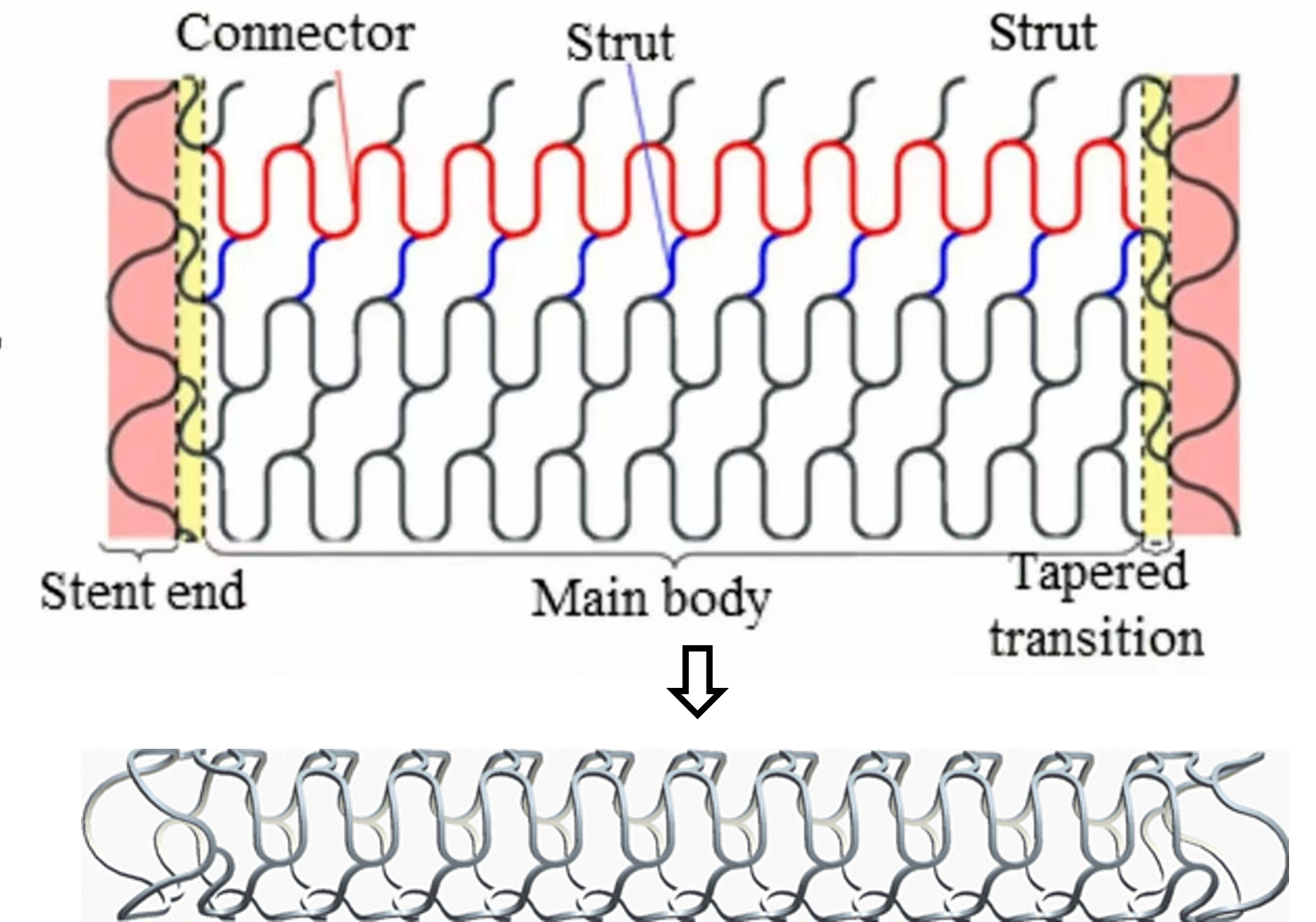
Figure 1. Non-modular stent structure: high-flexibility stent structure[38].
3.2. Optimization Design Based on Existing Stent Structures
Li et al. optimized the rib width, rib thickness, and curvature radius of the support unit curved parts of the Palmaz-Schatz stent and SV stent. After optimization, the dog-boning rate (a phenomenon where the diameter of the stent ends is larger than that of the middle part during expansion) during the expansion of the two stents was almost zero, proving that their expansion uniformity was improved, and the optimized stent has a higher fatigue life. Wu et al.[39-41] optimized the coronary stent Magic of Biotronik Company. The configuration of the stent support unit was slightly adjusted after optimization, causing the maximum stress value to be reduced during the expansion process, improving the degradation characteristics of the stent. Chen et al.[42] further optimized the stent based on the work, reduced the maximum stress value during the compression process, and improved its corrosion resistance. Hsiao et al.[43] optimized the width of the supporting unit, so that it gradually shortened from the end to the middle position, showing a taper. The optimized stent can concentrate stress during the expansion process from the bent part of the support unit to the linear part, improving the anti-fatigue characteristics of the coronary stent. García et al.[44] proposed a stent optimization scheme with variable radial stiffness. The stent is divided into three parts, so that the rib thickness of the two ends of the stent is smaller than the middle part, thereby reducing the radial stiffness of the vascular healthy area during the stent service. From the above research, we can see that the main objects of stent structure optimization are their rib width and rib thickness, and the side shows that the rib width and rib thickness are of great significance to the study of stent performance. However, the nature of the impact of rib width and rib thickness on the performance of the stent is not further clarified.
3.3. Comparison of Different Stent Structures
Italian scholar Petrini et al. conducted a comparative analysis of the bending behaviors between the Cordis BX-Velocity stent (CV) from Johnson & Johnson (USA) and the Sirius Carbostent (SV) from Sorin (Italy). The research results show that the flexibility of the CV model structure is better than that of SC, and the former has better independent deformation ability compared to the latter. Azaouzi et al.[45] compared the influence of four different connecting unit structures of coronary stents on their bending, torsion, and expansion behavior. The results show that the connecting unit structure mainly has a great impact on its bending and torsion, and the unsymmetrical N-shaped bridge structure shows good mechanical properties in both flexibility and torsion of the stent. Bobel et al.[46] compared the radial compression, bending flexibility, and axial compression of the three commercial thrombotic stent structures. The Absorb stent structure has the largest radial stiffness, the Zig-Zag stent structure has the best flexibility, and the Multilink stent structure has better axial compression. Hejazi et al.[47] compared the mechanical properties of four commercial thrombotic stents through experiments. Experimental results show that the Diamond stent and Braided stent have high radial stiffness, the Braided stent has the best overall compressive performance, and the Z stent has the best local compressive performance. Among the four types of stents, the Chevron stent demonstrated the poorest performance in terms of radial stiffness, as well as overall and local compressive resistance. The above study clarifies the impact of structure on the mechanical properties of the stent through comparative methods, but few researchers have explained it in-depth, which is not conducive to understanding stent design. Shi et al.[48] carried out numerical simulation research on the bending process of six different connecting unit stents. As shown in Figure 2, they analyzed the impact of the connection mode and shape of the connecting unit on the flexibility of the stent, and established a general mathematical model between the structural parameters and flexibility of the connecting unit, revealing the essence of the regular changes in the high stress zone of the stent connection unit during bending. The results show that the slashed "W"-shaped connecting unit stent has the best flexibility, and the linear part length and curvature radius of the curved part are positively correlated with the flexibility. Compared with the curvature radius of the curved part, the influence of the linear part length on flexibility is more significant.
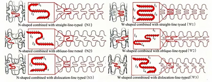
Figure 2. N 2D and 3D geometry of stent bridges[48].
4. Research on Key Mechanical Properties of Biodegradable Cerebrovascular Stents
Any vascular stent structure is designed to obtain the performance shown by the structure. The design parameters that meet the mechanical properties of the stent are often contradictory, and the final stent structural design is usually the result of highlighting the main performance and compromising other performances. Therefore, the primary goal of developing a new stent is to determine its main performance based on clinical application, and to master the current characterization and testing methods of this performance, so as to optimize the stent structure more targeted based on feedback.
The cerebrovascular pathways, especially the siphon segment of the internal carotid artery, can form a relatively sharp spatial angle[49]. On the one hand, this anatomical feature requires that the stent should have high flexibility to ensure that it can reach the lesion target, and the stent should also have a certain degree of flexibility to adapt to the complex arterial wall shape[1]. In addition, the stent needs to withstand pulsating loads from the blood vessel wall during service. Therefore, the development of biodegradable cerebrovascular stents is first concerned with flexibility, but its support must not be ignored.
4.1. Flexibility
Flexibility is an important characteristic of vascular stents, and it can characterize the axial bending characteristics of vascular stents[27, 50]. During the implantation process, the stent needs to pass through various tortuous blood vessel ducts and can adapt to various blood vessel shapes after dilatation at the lesion position[51]. If the stent is too poor, it will directly cause the stent to fail to reach the lesion position smoothly, resulting in the failure of the operation. In addition, flexibility is also related to the efficacy of the lesion blood vessels. If the performance is too poor, it will change the original morphology of the bent lesion blood vessels, that is, over-standing[52], which will directly cause damage to different degrees of blood vessels and increase postoperative complications.
The test methods for flexibility can be divided into four types, three-point bending method[5, 27, 53-55], four-point bending method[36, 56], cantilever beam bending method[33, 50], and vascular modeling method[52, 57-59]. The three-point bending method is the most commonly used method at present. One end of the stent is fixed, and a force (approximately 0.5 N) is applied axially at a position (approximately 10 mm) from the free end to induce bending, while recording the bending angle, recording the displacement of the loading component and the reaction force being received. The vascular modeling method simulates the implantation process of the stent. Two loading components simultaneously apply pressure to the stent, which is expected to avoid radial bending or local deformation of the stent during the three-point bending process[52]. The cantilever beam bending method is a common flexibility test method in the literature[33, 50]. One end of the stent is fixed. A force of approximately 0.5 N is applied axially at a position about 10 mm from the free end to induce bending. Simultaneously, the bending angle, displacement of the loading component, and the reaction force received are recorded. The vascular modeling method simulates the implantation process of the stent. The stent passes through a curved pipe made of silicone or glass at a constant speed under the action of traction or thrust, recording the magnitude of the traction or thrust when passing through the bend. This method greatly reduces the stent implantation process.
Except for the three-point bending method, there is currently no complete evaluation system for the other three methods to regulate their operation[27], resulting in some uncertainty or interference factors that affect the accuracy of the measurement results when they are used to test the flexibility of the stent. For example, in the four-point bending method, how to set the distance between the two staking points. In the cantilever beam bending method, how to determine the position of the staking point, and how to evaluate the impact of the relative slippage of the stent on the surface of the stent during bending. And how to eliminate the impact of friction and traction speed on the result generated by different stents in the vascular modeling method, etc.
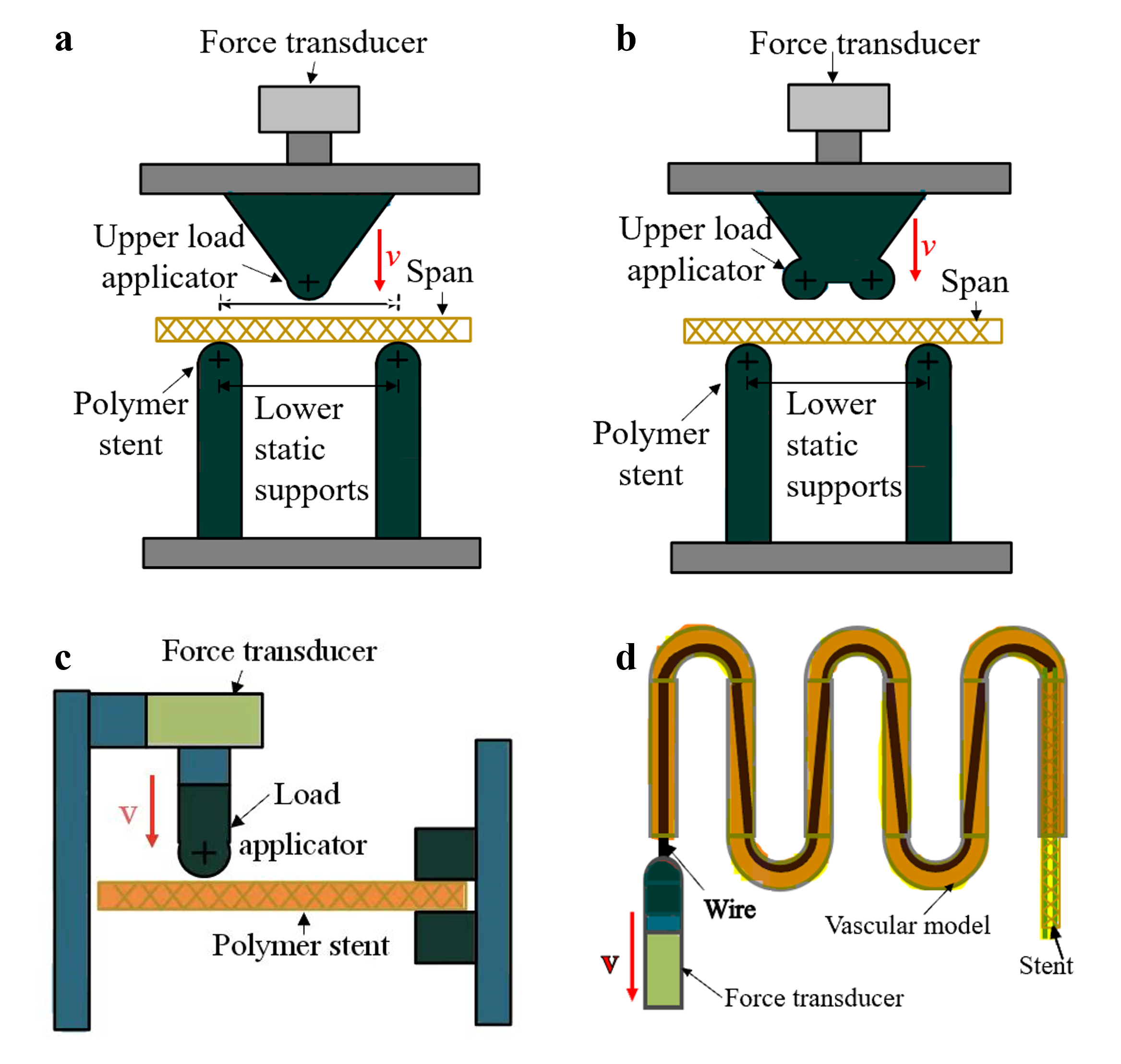
Figure 3. Schematics of different test method for stent flexibility[60]: (a) three-point bending, (b) four-point bending, (c) cantilever beam bending, and (d) vascular modeling.
Researchers usually use the finite element method to study the flexibility of the stent[61], which has the advantages of accurate calculations and complete information. Feng et al.[62] used the finite element method to compare the flexibility of the Palmaz-Schatz stent with V- and S-shaped connecting unit stents, and found that the shape of the connecting unit has a great influence on the flexibility of the stent. At the same time, the change in the flexibility of the stent after cyclic bending was studied. The results showed that cyclic bending increased the stiffness of the stent. Bobel et al.[46] used the finite element method to study the effect of the thickness of the stent rib on the flexibility of the three commercial stents, and showed that the flexibility of the stent can be significantly improved by reducing the thickness of the stent ribs. Shen et al.[63] studied the impact of the number of connecting units on the flexibility of the stent, and the simulation results showed that the reduction of the number of connecting units greatly improved the flexibility of the stent.
4.2. Radial Support
The support of the stent is a ability of the vascular stent to resist the contraction of the vascular wall[64], allowing the stent to keep the blood vessels open without collapse on its own after angioplasty. Different diseases have different needs for stent support, such as cardiovascular thrombosis and cerebral aneurysm diseases. As shown in Figure 3(a), the thrombus stent requires greater support to support the thrombus in the blood vessels to ensure the smoothness of blood flow at the lesion location[36, 65]. However, the need for supportiveness of the brain aneurysm stent is relatively small. After release, the cerebral aneurysm stent can be effectively fixed to the lesion site and will not be affected by pulsation, skeletal muscle interactions caused by patient activities, and various external influences. It can also ensure that the curved stent does not suffer from large cross-sectional loss or collapse during service[66], and the radial force required for these requirements is less than the radial force required to support the thrombus, as shown in Figure 3 (b). In addition, the support during the stent service should not be too large. Excessive support usually means that the stent has been over-drawn, which can easily cause blood vessel damage. The ideal state is that during the service of the stent, the support of the stent just keeps the inner wall of the stent in line with the inner wall of the blood vessel.
The supportability can be characterized by testing the radial stiffness of the stent. There are generally two methods for measuring the radial stiffness of the stent, the radial loading method[42, 67, 68] and the planar compression method[44, 54, 67, 69-71]. Radial loading can better simulate the service environment of the stent, but special instruments are required, such as iris coilers and radial force testers[42, 72]. Figure 4 (a) is a schematic diagram of the radial loading method. Compared with the radial loading method, the planar compression method is a relatively common method of radial stiffness testing of stents. The main reason is that this method can be tested using ordinary experimental equipment. The principle of the planar compression method is shown in Figure 4 (b). The stent is placed between the two plates, and the upper plate is compressed by applying displacement. In the process of compressing the stent, the displacement of the upper plate and the reaction force can be obtained through the sensor, and the reaction force is used to describe the radial stiffness of the stent.
The initial development of vascular stents was to restore the vascular diameter of the lesion site and ensure smooth blood flow. At present, a large number of literatures[67, 70, 73-75] have studied the impact of the structural parameters of stents on support. As the research deepens, stents are used to treat or assist in the treatment of other vascular diseases, especially cerebrovascular diseases. According to the structural characteristics of cerebrovascular systems, the stent has been redesigned, the balance between various mechanical properties has been redefined, and flexibility has gradually been valued by researchers. However, compared with support, the study of flexibility is still relatively lagging. For example, researchers have tried to crack the law of the influence of structural parameters on radial support through mathematical mode[69, 74], but the mathematical relationship between scaffold structural parameters and their bending flexibility is still in the exploration stage. Although some researchers have conducted preliminary research on the bending behavior of stents using the finite element method, these studies mainly focus on the comparison of the flexibility of stents of different structures. The mechanism of the impact of stent connection unit structure on bending behavior has not been fully analyzed, and it is impossible to further guide the design of new biodegradable metal cerebrovascular systems. And most studies ignore the analysis of bending deformation patterns during stent service, which also limits the optimized design of stent structure in clinical applications.
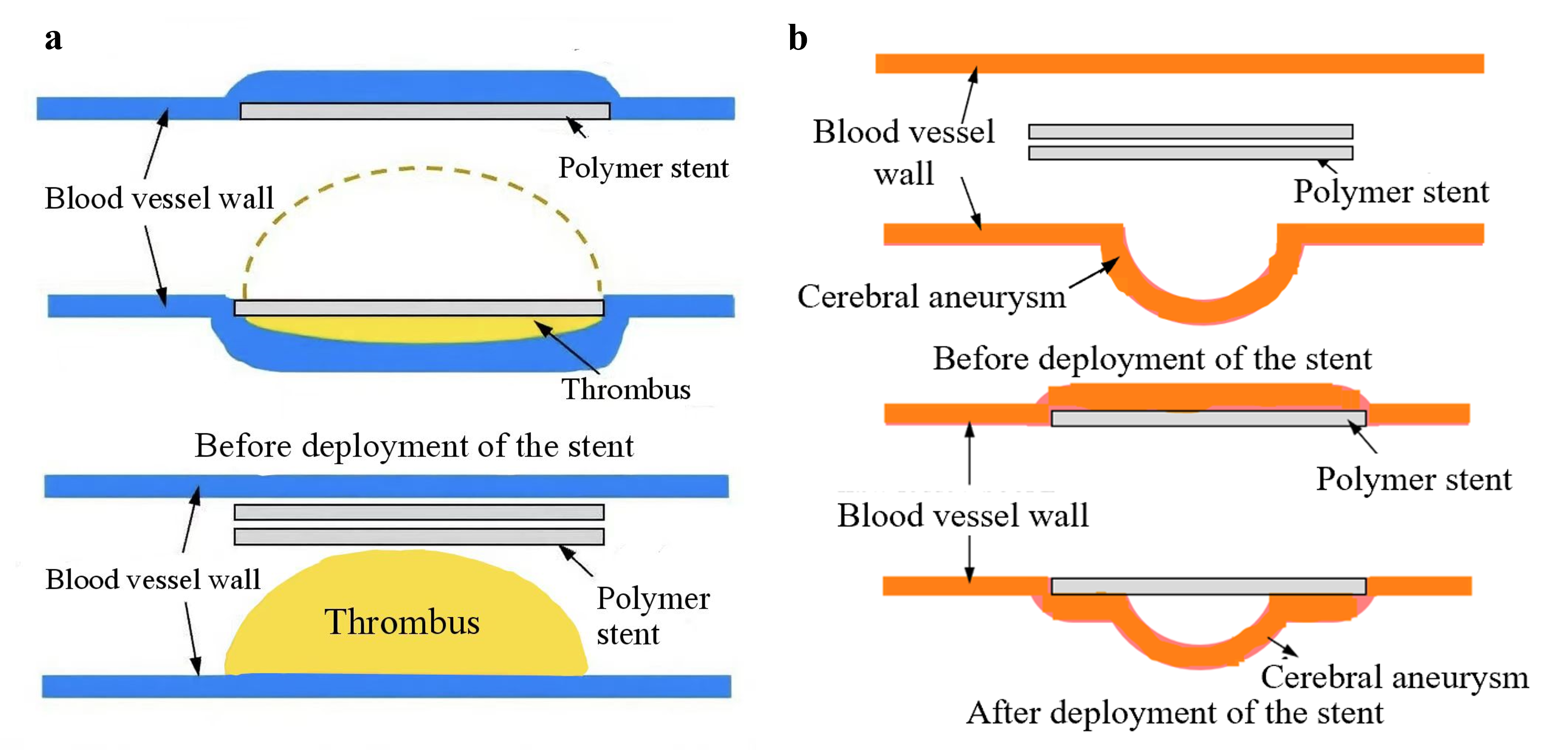
Figure 4. Radial stiffness requirements for different stents[60]: (a) thrombus stents and (b) cerebral aneurysm stents
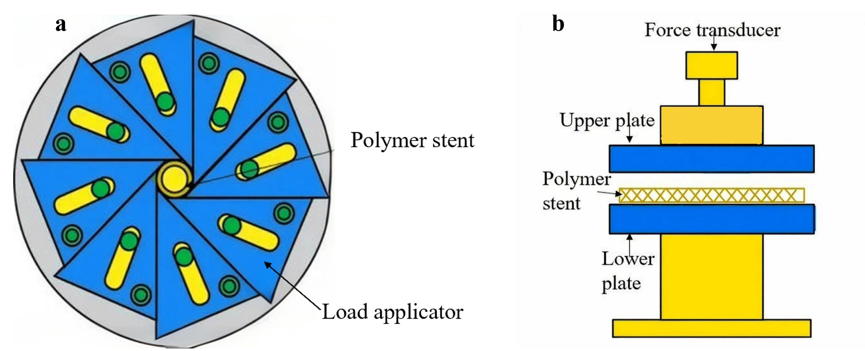
Figure 5. Test method for radial stiffness of stents[60]: (a) radial loading method and(b) planar compression mentod.
4.3. Other Mechanical Properties
In addition to flexibility and support, other common mechanical properties of vascular stents include grip, radial retraction, and axial shortening[76]. The grip of the stent is the ability of the stent to reach a specified diameter without loss by radial compression, which ensures that the stent is effectively fixed to the balloon surface of the delivery system[43, 77]. Radial retraction refers to the change in the diameter and size of the stent from the end of balloon expansion to the unloading process, which is related to the fixation effectiveness of the stent at the lesion position[42, 78]. Axial shortening indicates the change in length of the stent after it expands from the grip state to the nominal diameter, which is related to the positioning of the stent in the lesion vessel after its release[42]. During the stent design stage, according to clinical needs, as much as possible, other performance influencing factors should be considered comprehensively on the basis of ensuring the main performance of the stent, and the relationship between the various performances should be balanced, so that the stent is more conducive to clinical treatment.
5. Discussion and Conclusion
Through the above analysis, we can see that domestic and foreign researchers have conducted a series of research on biodegradable metal vascular stents and have achieved certain research results. However, existing stents will still be limited in clinical application due to mechanical properties and other reasons when treating cerebrovascular diseases, which are mainly manifested in the following points
The bending mechanism of the stent structure is not yet clear. At present, the research on the flexibility of vascular stents focuses on the influence of the thickness, shape and number of stent connection unit ribs on flexibility, and relatively few studies have been conducted on the impact of connection methods on them. The research content also focuses on comparing the advantages and disadvantages of stent flexibility in different structures. The research on the mechanism of its action is not in-depth enough, and most studies ignore the impact of stents on hemodynamics when the stent is deformed, and there is a lack of discussion on the bending deformation of stents.
The existing vascular stent structure has structural defects in the treatment of cerebrovascular diseases. Research on the design of biodegradable metal stent structures is mainly aimed at the treatment of cardiovascular diseases. The original intention of the research is to enhance the radial support of the stent or optimize the expansion behavior of the stent. However, the walls of cerebrovascular vessels are thin and the paths are tortuous, and the anatomical characteristics are completely different from those of the cardiovascular system. Research on its stents should focus more on flexibility and bending behavior. Therefore, the existing stent structure is not suitable for the treatment of cerebrovascular diseases due to its insufficient flexibility. The traditional modular stent structure as the mainstream design of cardiovascular stents has been recognized by researchers, resulting in the fact that the existing stent structure design is mostly limited to this design method, and there is no major breakthrough in the prototype design of cerebrovascular stents.
Author Contributions
L.H. and W.S. is responsible for conceptualization and methodology design, and drafts the original manuscript, establishing the core framework of the review. J.L., J.W, Y.D. and H.W. undertakes investigations, provides resources, and conducts data visualization to present the research results intuitively. F.J., G.W., B.G., and M.Z. are in charge of reviewing and editing the manuscript while supervising the project to control the overall quality and progress. W.S. is responsible for funding acquisition and project administration, providing guarantees for the smooth progress of the research.
Acknowledgements
This research was funded by Shandong Provincial Postdoctoral Science Foundation (SDCX-ZG 202400013)
References
-
Y. Zh, H. Zhang, Y. Zhang, H. Wu, L. Wei, G. Zhou, Y. Zhang, L. Deng, Y. Cheng, M. Li, H. Santos, W. Cui. "Endovascular metal devices for the treatment of cerebrovascular diseases". Adv. Mater, 2019, 31, 8, 1805452.
-
Q. Huang, P. Yang. "Chinese Expert Consensus on Endovascular Intervention for Intracranial Aneurysms(2013)". Chin J of Cerebrovasc Dis, 2013, 10, 11, 606-16.
-
D. Jiang, C. Qian. "Advances in Endovascular Intervention for Intracranial Aneurysms.". Zhejiang Med J, 2019, 41, 19, 2033-40.
-
Y. Ito, I. Cho, Y. Sakai, K. Iwano. "CFD study on the efficacy of flow diverter stent placement for cerebral aneurysms". JA FM, 2021, 14, 5, 1547-58.
-
C. Rutledge, F. Baranosk, S. Catapano, T. Lawton, F. Spetzler. "Microsurgical treatment of cerebral aneurysms". World Neurosurg, 2022, 159250-8.
-
X. Lv, J. Zang. "Comparative Efficacy of Craniotomy Clipping versus Endovascular Coiling for Ruptured Intracranial Aneurysms". Zhejiang J of Traumatic Surg, 2018, 23, 5, 916-7.
-
M. Kyle, E. Hector, T. Julia, W. Mack, J. Mocco, F. Albuquerque, F. Andrew, M. Mokin, I. Linfante, Q. Stacey, A. John, A. Joshua. "A survey of intracranial aneurysm treatment practices among United States physicians". JNIS, 2018, 10, 1, 44-9.
-
F. Li, K. Wu, J. Zhao, G. Li. "Vascular Stents and Their Development Trends in the Treatment of Aneurysms". CJTER, 2021, 25, 34, 5561-9.
-
X. Li. "Implantation of Vascular Stents for the Treatment of Atherosclerotic Arterial Stenosis in Canine Models", 2023.
-
S. Wang, H. Zhang, M. Zhu, K. Shu. "Strategies for Cerebral Revascularization in Complex Intracranial Aneurysms". The 18th Annual Conference of the Chinese Congress of Neurological Surgeons, Xiamen, 2024.
-
C. Qin, J. Liu. "Diagnosis and treatment of subarachnoid hemorrhage". Chin. J. Neurol., 2020, 53, 10, 814-8.
-
G. Guido, V. Fernando, D. Jacques, D. Gary. "Electrothrombosis of saccular aneurysms via endovascular approach". J. Neurosurgery, 1991, 758-14.
-
R. Higashida, W. Smith, D. Gress, R. Urwin, C. Dowd, P. Balousek, V. Halbach. "Intravascular stent and endovascular coil placement for a ruptured fusiform aneurysm of the basilar artery - Case report and review of the literature". J. Neurosurgery, 1997, 87, 6, 944-9.
-
M. Azaouzi, A. Makradi, J. Petit, S. Belouettar, O. Polit. "On the numerical investigation of cardiovascular balloon-expandable stent using finite element method". Comput. Mater. Sci., 2013, 79326-35.
-
S. Cho, W. Jo, Y. Jo, H. Ku, C. Jung, H. Deok. "Bench-top comparison of physical properties of 4 commercially-available self-expanding intracranial stents". Neurointerv., 2017, 12, 1,31-9.
-
M. Cabrer, C. Oomens, F. Baaijens. "Understanding the requirements of self-expandable stents for heart valve replacement: radial force, hoop force and equilibrium". JMBBM, 2017, 68252-64.
-
A. Mohamed, M. Ahmed, B. Salim. "Deployment of a self-expanding stent inside an artery: a finite element analysis". Mater. Des., 2012, 41410-20.
-
Q. Shao, L. Li, T. Li, K. Chang, L. Zhu, J. Xue, Z. Wang, G. Zhang, Q. Zhang, Q. Wu. "Preliminary application of Neuroform Atlas stent-assisted coiling in the treatment of intracranial wide-necked aneurysms". Chin. J. Neurosurg., 2022, 38, 01, 59-64.
-
S. Sun, C. Xu, P. Wu, M. Li, S. Xu, C. Wang, X. Liu, Y. Ling, Huaizhang Shi. "Intracranial angioplasty with Enterprise stent for intracranial atherosclerotic stenosis: a single-center experience and a systematic review". Biomed Res. Int., 2021, 20216645500.
-
H. Tang, S. Li, F. Xu, C. Shang, Z. Lu, Z. Zeng, H. Yin, Q. Zuo, Q. Li, Q. Huang, J. Liu. "Low-profile LEO baby stents using dual stenting technique in treating complex intracranial aneurysms located in small artery: initial and mid-term outcome". J Clin Neurosci, 2022, 98109-14.
-
R. Zang, J. Wang, X. Cao, Z. Du, X. Liu, B. Lv, Z. Wang, B. Li, S. Yu. "Application of Neuroform EZ stent and Solitaire AB stent in the treatment of severe intracranial atherosclerotic stenosis.". Chin. J. An. at. Clin., 2021, 26, 04, 391-6.
-
W. Jiang, W. Zhao, T. Zhou, L. Wang, T. Qiu. "A review on manufacturing and post-processing technology of vascular stents". MICROMACHINES-BASEL, 2022, 13, 1, 140.
-
A. Kalra, H. Rehman, S. Khera, B. Thyagarajan, L. Deepak, S. Neal, W. Robert. "New-generation coronary stents: current data and future directions". CURR ATHEROSCLER REP, 2017, 19, 3, 14.
-
H. Zhao, C. Li. "Optimization Design and Mechanical Performance Analysis of Bioabsorbable Polymeric Vascular Stents". J. Liaoning Univ. of Technol., 2024, 44, 04, 234-7+44.
-
Y. Yang, Z. Zhang, J. Wang, K, Fu, D. Li, H. He, C. Shu. "Research Progress in Degradable Metal Vascular Stents". JCSU, 2024, 49, 11, 1861-8.
-
R.Zhou."Research on 3D printing technology of biodegradable polymer vascular stent", 2019.
-
ASTM Committee F04 on Medical and Surgical Materials and Devices. "Standard guide for three-point bending of balloon expandable vascular stents and stent systems". 2014, F2606−08.
-
China Food and Drug Administration. "Standard guide for characterization and presentation of the dimensional attributes of vascular stents". 2008, YY/T 0693—2008.
-
Y. Wei. "Structural Design and Mechanical Property Test of Biodegradable Polymeric Stent"; DUT, 2020.
-
S. Dun. "Study on Micro Injection Molding Technology of Biodegradable Stent." DUT, 2013.
-
C. Wang, L. Zhang, Y. Fang, W. Sun. "Design, characterization, and 3D printing of cardiovascular stents with zero Poisson's ratio in longitudinal deformation". ENGINEERING-PRC, 2021, 7,7, 979-90.
-
K. Song, Y. Bi, H. Zhao, T. Wu, F. Xu, G. Zhao. "Structural optimization and finite element analysis of poly-l-lactide acid coronary stent with improved radial strength and acute recoil rate". J. Biomed. Mater. Res. Part B Appl. Biomater., 2020, 108,7, 2754-64.
-
S. Yasuhiro, T. Tetsuya, T. Satoshi, T. Kazuo. "Mechanical design of an intracranial stent for treating cerebral aneurysms". Med. Eng. Phys., 2010, 32, 9, 1015-24.
-
S. De Bock, F. Iannaccone, G. De Santis, M. De Beule, P. Mortier, B. Verhegghe, P. Segers. "Our capricious vessels: The influence of stent design and vessel geometry on the mechanics of intracranial aneurysm stent deployment". J. Biomech., 2012, 45, 8, 1353-9.
-
N. Ebrahimi, B. Claus, C. Lee, A. Biondi. "Stent conformity in curved vascular models with simulated aneurysm necks using flat-panel CT: an in vitro study". AJNR, 2007, 28, 5, 823-9.
-
X. Ruan, W. Yuan, Y. Hu, J. Li, W. Wu, R. Xia. "Chiral constrained stent: effect of structural design on the mechanical and intravascular stent deployment performances". Mech. Mater., 2020, 148103509.
-
C. Singh, X. Wang. "A biomechanically optimized knitted stent using a bio-inspired design approach". Text. Res. J., 2016, 86, 4, 380-92.
-
W. Shi, C. Zhang, A. Xie, K. Mitchell, Y. Jin, D. Zhao. "Development of a Computational Framework for the Evaluation of Biodegradable Cerebral Stents With Enhanced Bending Performance". J. Med. Devices, 2023, 17, 1, 1-14.
-
W. Wu, L. Petrini, D. Gastaldi, T. Villa, M. Vedani, E. Lesma, B. Previtali, F. Migliavacca. "Finite element shape optimization for biodegradable magnesium alloy stents". Ann. Biomed. Eng., 2010, 38, 9, 2829-40.
-
W. Wu, D. Gastaldi, K. Yang, L. Tan, L. Petrini, F. Migliavacca. "Finite element analyses for design evaluation of biodegradable magnesium alloy stents in arterial vessels". Mater. Sci. Eng. B, 2011, 176, 20, 1733-40.
-
W. Wu, S. Chen, D. Gastaldi, L. Petrini, D. Mantovani, K. Yang, L. Tan, F. Migliavacca. "Experimental data confirm numerical modeling of the degradation process of magnesium alloys stents". Acta Biomater, 2013, 9, 10, 8730-9.
-
C. Chen, J. Chen, W. Wu, Y. Shi, L. Jin, L. Petrini, L. Shen, G. Yuan, W. Ding, J. Ge, R. Edelman, F. Migliavacca. "In vivo and in vitro evaluation of a biodegradable magnesium vascular stent designed by shape optimization strategy". Biomaterials, 2019, 221119414.
-
M. Hao, L. Wu, M. Yin, C. Lin, H. Chen. "Quintupling fatigue resistance of intravascular stents via a simple design concept". Computat. Mater. Sci., 2014, 8657-63.
-
A. Garcia, E. Pena, M. Martinez. "Influence of geometrical parameters on radial force during self-expanding stent deployment. Application for a variable radial stiffness stent". J. Mech. Behav. Biomed. Mater., 2012, 10166-75.
-
M. Azaouzi, A. Makradi, S. Belouettar. "Numerical investigations of the structural behavior of a balloon expandable stent design using finite element method". Comput. Mater. Sci., 2013, 7254-61.
-
C. Bobel, S. Petisco, J. Sarasua, W. Wang, P. Mchugh. "Computational bench testing to evaluate the short-term mechanical performance of a polymeric stent". Cardiovasc. Eng. Techn., 2015, 6, 4, 519-32.
-
M. Hejazi, F. Sassani, J. Gagnon, Y. Hsiang, S. Phani. "Deformation mechanics of self-expanding venous stents: modelling and experiments". J. Biomech., 2021, 120110333.
-
W. Shi, H. Li, T. Zhu, Y. Jin, H. Wang, J. Yang, D. Zhao. "Study on the bending behavior of biodegradable metal cerebral vascular stents using finite element analysis". J. Biomech., 2020, 108109856.
-
X.Yang, J.Tian, S.Liu, J.Tian, Y.Zhao, C.Tian. "Study on the anatomical characteristics of internal carotid artery siphon in patients with intracranial aneurysm and its correlation with aneurysm rupture". Chin J New Clin Med, 2023, 16, 04, 379-83.
-
Y. Guan, J. Lin, Z. Dong, L. Wang. "Comparative study of the effect of structural parameters on the flexibility of endovascular stent grafts". Adv. Mater. Sci. Eng., 2018, 20181-10.
-
Z.Ma, M.Bai, S.Li. "Optimal design of vascular stents based on complimentation and strength reliability analysis". Mod. Instrum. Med. T., 2024, 30, 02, 23-8.
-
N. Tanaka, J. Martin, K. Tokunaga, T. Abe, Y. Uchiyama, N. Hayabuchi, J. Berkefeld, D. Rüfenacht. "Conformity of carotid stents with vascular anatomy evaluation in carotid models". AJNR Am J Neuroradiol, 2004, 25, 4, 604-7.
-
S. Lee, H. Jo, K. Lim, D. Lim, S. Lee, J. Lee, W. Kim, M. Jeong, J. Lim, I. Kwon, Y. Jung, J. Park, S. Park. "Heparin coating on 3D printed poly (l-lactic acid) biodegradable cardiovascular stent via mild surface modification approach for coronary artery implantation". Chem. Eng. J., 2019, 378122116.
-
J. Singh, P. Pandey, T. Kau, N. Singh. "A comparative analysis of solvent cast 3D printed carbonyl iron powder reinforced polycaprolactone polymeric stents for intravascular applications". J. Biomed. Mater. Research Part B: Appl. Biomater., 2021, 109, 9, 1344-59.
-
K. Somszor, O. Bas, F. Karimi, T. Shabab, N. Saidy, A. O’Connor, A. Ellis, D. Hutmacher, D. Heath. "Personalized, mechanically strong, and biodegradable coronary artery stents via melt electrowriting". ACS Macro Lett., 2020, 9, 12, 1732-9.
-
M. Koji, S. Takashi. "Effects of stent structure on stent flexibility measurements". ANN BIOMED ENG, 2005, 33, 6, 733-42.
-
J. Liu, B. Zheng, P. Wang, X. Wang, B. Zhang, Q. Shi, T. Xi, M. Chen, S. Guan. "Enhanced in vitro and in vivo performance of Mg-Zn-Y-Nd alloy achieved with APTES pretreatment for drug-eluting vascular stent application". ACS Appl. Mater. Interfaces, 2016, 8, 28, 17842-58.
-
R. du Mesnil, B. Yan, F. Zanella, D. Rüfenacht, J. Berkefeld. "Conformability of balloon-expandable stents to the carotid siphon: an in vitro study". AJNR, 2006, 27, 2, 324-6.
-
R. Rieu, P. Barragan, V. Garitey, R. Pierre, J. Fuseri, P. Commeau, J. Sainsous. "Assessment of the trackability, flexibility, and conformability of coronary stents: a comparative analysis". Catheter. Cardiovasc. Interv., 2003, 59, 4, 496-503.
-
W. Hua, W. Shi, K. Mitchell, L. Raymond, R. Coulter, D. Zhao, Y. Jin. "3D printing of biodegradable polymer vascular stents: a review". CJME: AMF, 2022, 1, 2, 100020.
-
L.Liu, Y.Dong, H.Ding, Y.Lin, H.Wang. "Application of finite element method in evaluation of biomechanical performance of vascular stents". Mech. Des. Res., 2015, 31, 01, 35-8.
-
F. Ju, Z. Xia, C. Zhou. "Repeated unit cell (RUC) approach for pure bending analysis of coronary stents". Comput Methods Biomech Biomed Engin, 2008, 11, 4, 419-31.
-
X. Shen, Q. Yong, S. Ji, Z. Xie, H. Zhu. "Flexibility behavior of coronary stents: the role of linker investigated with numerical simulation". J. Mech. Med. Biol., 2017, 17, 08, 1750112.
-
R. Pauck, B. Reddy. "Computational analysis of the radial mechanical performance of PLLA coronary artery stents". Med. Eng. Phys, 2015, 37, 1, 7-12.
-
S. Shi, M. Cui, F. Sun, K. Zhu. "An innovative solvent‐responsive coiling–expanding stent". Adv. Mater., 2021, 33, 32, 2101005.
-
Administration National Medical Products. "Guide for radial loading of balloon expandable and self-expanding vascular stents.", 2019.
-
Z. Wu, J. Zhao, W. Wu, P. Wang, B. Wang, G. Li, S. Zhang. "Radial compressive property and the proof-of-concept study for realizing self-expansion of 3D printing polylactic acid vascular stents with negative Poisson's ratio structure". Materials, 2018, 11,8, 1357.
-
S. María, B. Sanders, J. Olga, A. Mol, W. Cees, P. Frank. "Computationally designed 3D printed self-expandable polymer stents with biodegradation capacity for minimally invasive heart valve Implantation: a proof-of-concept study". 3D Print. Addit. Manuf., 2017, 4, 1, 19-29.
-
R. Kandi, M. Pulak. "Statistical modelling and optimization of print quality and mechanical properties of customized tubular scaffolds fabricated using solvent-based extrusion 3D printing process". Proc. Inst. Mech. Eng. Part H: J. Eng. Med., 2021, 235, 12, 1421-38.
-
Y. Wei, M. Wang, D. Zhao, H. Li, Y. Jin. "Structural design of mechanical property for biodegradable polymeric stent". Adv. Mater. Sci. Eng., 2019, 20191-14.
-
T. Qiu, W. Jiang, P. Yan, L. Jiao, X. Wang. "Development of 3D-printed sulfated chitosan modified bioresorbable stents for coronary artery disease". Front. Bioeng. Biotechnol., 2020, 8462.
-
ISO/TC 150/SC 2 Cardiovascular implants and extracorporeal systems. "Cardiovascular implants — Endovascular devices — Part 2: Vascular stents". 2012, ISO 25539-2.
-
K. Jang, T. Kang, K. Lee, C. Kim, T. Kim. "The effect of change in width on stress distribution along the curved segments of stents". J. Mech. Sci. Technol., 2010, 24, 6, 1265-71.
-
J. Yang, N. Huang. "Mechanical formula for the plastic limit pressure of stent during expansion". Acta Mechanica Sinica, 2009, 25, 6, 795-801.
-
J. Hiba, M. Sofiene, C. Nabil, H. Frederic, A. Ben. "Elastic recovery of polymeric braided stents under cyclic loading: Preliminary assessment". J. Mech. Behav. Biomed. Mater., 2019, 98131-6.
-
L.Yang, L.Sheng. "Structural design and mechanical properties analysis of Ni-Ti alloy vascular stent". JMRA, 2025, 38, 01, 14-20.
-
C. Pan, Y. Han, J. Lu. "Structural design of vascular stents: a review". MICROMACHINES-BASEL, 2021, 12, 7, 770.
-
Administration China Food and Drug. "Cardiovascular implants Endovascular devices—Part 2: Vascular stents.", 2016.